Arxiu de la categoria: CONTENTS

The extended phenotype: genetics beyond the body

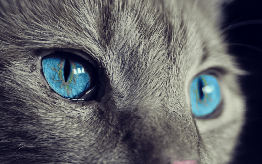
Genes determine our eye color, height, development throughout life and even our behavior. All living beings have a set of genes that, when expressed, manifest themselves in a more or less explicit way in their body, modeling it and giving it a wide diversity of traits and functions. However, is it possible that the expression of some genes has effects beyond the body itself?

Discover some basic ideas about the extended phenotype theory.

The extended phenotype: genetics beyond the body

First of all, let’s talk about two basic, but not less important, concepts that will help you to understand the extended phenotype theory: genotype and phenotype.

Genotype

Genotype is the collection of genes or the genetic information that a particular organism possesses in the form of DNA. It can also refer to the two alleles of a gene (or alternative forms of a gene) inherited by an organism from its parents, one per parent.

The genetic information that a particular organism possesses in the form of DNA constitutes its genotype. Public domain image.

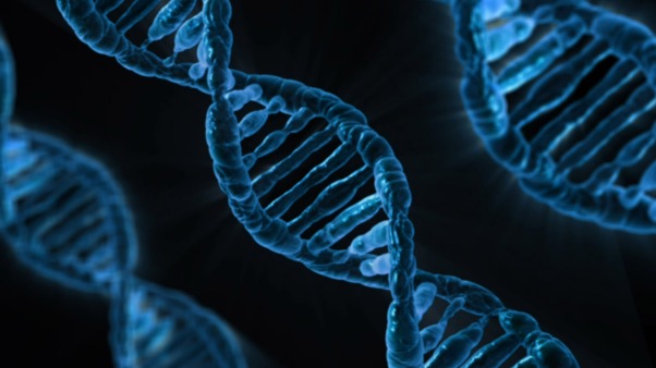
It should not be confused with the genome: the genome is the set of genes conforming the DNA that a species has without considering its diversity (polymorphisms) among individuals, whereas the genotype does contemplate these variations. For example: the human genome (of the whole species Homo sapiens sapiens) and the genotype of a single person (the collection or set of genes and their variations in an individual).

Phenotype

The genotype, or at least a part of it, expresses inside an organism thus contributing to its observable traits. This expression takes place when the information encoded in the DNA traduces to synthetize proteins or RNA molecules, the precursor to proteins. The set of observable traits expressed in an organism through the expression of its genotype is called phenotype.

Eye color (phenotype) is determined by the expression of a set of genes within an organism (genotype). Picture by cocoparisienne on Pixabay (public domain).

However, genes are not always everything when defining the characteristics of an organism: the environment can also influence its expression. Thus, a more complete definition of phenotype would be the set of attributes that are manifested in an organism as the sum of its genes and the environmental pressures. Some genes only express a specific phenotype given certain environmental conditions.

The extended phenotype theory

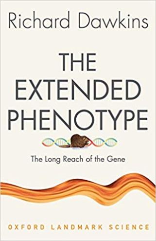
The concept of extended phenotype was coined by Richard Dawkins in his book “The Extended Phenotype” (1982). Dawkins became famous after the publication of what would be his most controversial work, “The Selfish Gene” (1976), which was a precursor to his theory of the extended phenotype.

In the words of Dawkins himself, an extended phenotype is one that is not limited to the individual body in which a gene is housed; that is, it includes “all the effects that a gene causes on the world.” Thus, a gene can influence the environment in which an organism lives through the behavior of that organism.

Dawkins also considers that a phenotype that goes beyond the organism itself could influence the behavior of other organisms around it, thus benefiting all of them or only one… and not necessarily the organism that expresses the phenotype. This would lead to strange a priori scenarios such as, for example, that the phenotype of an organism was advantageous for a parasite which afflicts it rather than for itself. This idea is summed up in what Dawkins calls the ‘Central Theorem of the Extended Phenotype’: ‘An animal’s behaviour tends to maximize the survival of the genes ‘for’ that behaviour, whether or not those genes happen to be in the body of the particular animal performing it’.

A complex idea, isn’t it? However, it makes sense if we take into account the basic premise from which Dawkins starts, which addresses in his work ‘The selfish gene’: the basic units of evolution and the only elements on which natural selection acts, beyond individuals and populations, are genes. So, organisms’ bodies are mere ‘survival machines’ improved to ensure the perpetuation of genes.

Examples of extended phenotype

Perhaps all these concepts seem very complicated, but you will understand them better with some examples. According to Dawkins, there exist three main types of extended phenotype.

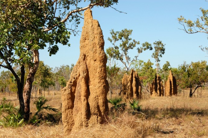
1) Animal architecture

Beavers build dams and modify their surroundings, in the same way that a termite colony builds a termite mound and alters the land as part of their way of life.

Dam built by beavers. Picture by Hugo.arg (CC 4.0)
Termite mounds in Autralia. Public domain image.

On the other hand, protective cases that caddisflies build around them from material available in the environment improve their survival.

Caddisfly larva inside its protective case made up of vegetal material. Picture by Matt Reinbold (CC 2.0)

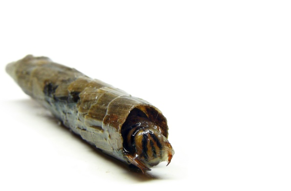
These are all examples of the simplest type of extended phenotype: the animal architecture. The phenotype is, in this case, a physical or material expression of the animal’s behavior that improves the survival of the genes that express this behavior.

2) Parasite manipulation of host behavior

In this type of extended phenotype, the parasite expresses genes that control the behavior of its host. In other words, the parasite genotype manipulates the phenotype (in this case, the behavior) of the host.

A classic example is that of crickets being controlled by nematomorphs or gordiaceae, a group of parasitoid ‘worms’ commonly known as hair worms, as explained in this video:

To sum up: larvae of hair worms develop inside aquatic hosts, such as larvae of mayflies. Once mayflies undergoe metamorphosis and reach adulthood, they fly to dry land, where they die; and it is at this point that crickets enter the scene: an adult cricket feeds on the remains of mayflies and acquires the hair worm larvae, which develop inside the cricket by feeding on its body fat. Adult worms must return to the aquatic environment to complete their life cycle, so they will control the cricket’s brain to ‘force’ it to find a water source and drop in. Once in the water, the worms leave the body of the cricket behind, which drowns.

Other examples: female mosquitoes carrying the protozoan that causes malaria (Plasmodium), which makes female mosquitoes (Anopheles) to feel more attracted to human breath than uninfected ones, and gall induced by several insects on different host plants, such as cynipids (microwasps).

3) Action at a distance

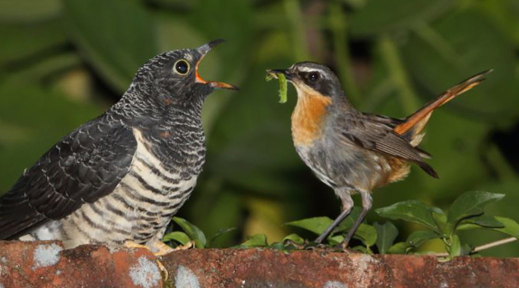
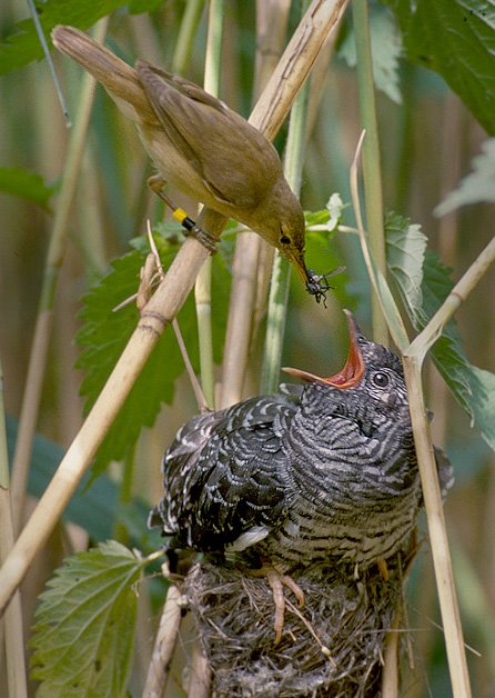
A recurring example of this type of extended phenotype is the manipulation of the host’s behavior by cuckoo chicks (group of birds of the Cuculidae family). Many species of cuckoo, such as the common cuckoo (Cuculus canorus), lay their eggs in the nests of other birds for them to raise in their place; also, cuckoo chicks beat off the competition by getting rid of the eggs of the other species.

Look how the cuckoo chick gets rid of the eggs of reed warbler (Acrocephalus scirpaceus)!

In this case of parasitism, the chick is not physically associated with the host but, nevertheless, influences the expression of its behavioral phenotype.

Reed warbler feeding a common cuckoo chick. Picture by Per Harald Olsen (CC 3.0).

.            .            .

There are more examples and studies about this concept. If you are very interested in the subject, I strongly recommend you to read ‘The selfish gene’ (always critical and from an open minded perspective). Furthermore, if you have good notions of biology, I encourage you to read ‘The extended phenotype’.

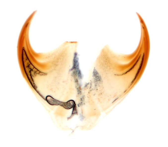
Main picture: Alandmanson/Wikimedia Commons (CC BY-SA 4.0)

The Asian giant hornet (Vespa mandarinia): What do we know about it?

Among the numerous exotic invasive organisms that have reached Europe and America, Asian wasps and hornets are some of the most commented on mass media, social networks and naturalistic forums. The Asian hornet (Vespa velutina) got Europe and, posteriorly, the Iberian Peninsula, becoming one of the greatest headaches for beekeepers and administrations as it is a very insatiable species. However, there exists an insect that concerns Westerner beekeepers even more than the Asian hornet: the Asian giant hornet (Vespa mandarinia).

What do we know about this species? Is it true is has been found in The West or is this a mere unfounded rumour? Keep reading to learn some more.

The Asian giant hornet (Vespa mandarinia): What do we know about it?

During my recent travel to Japan, I met face to face for the first time with one of the most amazing insects: the Asian giant hornet (Vespa mandarinia). Meeting this organism really inspired me to write this post.

The Asian giant hornet (Vespa mandarinia) is a hymenopteran native to the East and Southeast of Asia especially abundant in rural landscapes of Japan. Until recently, it was considered that the Japanese giant hornets belonged to an independent variety or subspecies (Vespa mandarinia japonica); however, this category is currently invalid.

Among the ‘true hornets’ (species belonging to the Vespa genus), the Asian giant hornet is the biggest worldwide. Workers of this species span between 3.5 to 4.0 cm long, whereas queens can reach a length between 5.0 to 6.0 cm, even more in some cases, and a wingspan of 3.5 to 7.5 cm depending on the specimen. A monster compared to the Asian hornet (Vespa velutina), which has a body length between 2.0 and 3.0 cm (3.5 in queens).

Vespa mandarinia Natural Museum of Natural Science Tokyo
Specimen of Vespa mandarinia (left) deposited in the main exhibition of the National Museum of Natural History of Tokyo, Japan. Picture by Irene Lobato Vila.

In fact, in Japan this species is commonly known as オオスズメバチ (oosuzumebachi), which can be translated as ‘sparrow wasp’.

How can we distinguish it from other related species?

The Asian giant hornet is easily recognizable and is distinguished from other Vespa species by its large size, as well as by having an orangish yellow head that can be seen even when the organism is in motion (and that differs from the rest of the body, which is darker), a well-developed clypeus and a very wide face seen from the front.

Face of Vespa mandarinia. Modified from the original picture took by Gary Alpert, CC 3.0.

In addition, and unlike the Asian hornet (V. velutina), it has darker legs (yellow in V. velutina) and the abdomen or metasoma with alternate yellow and black stripes (abdomen almost black, with the fourth segment yellow, in V. velutina).

Vespa mandarinia male
Vespa mandarinia. Picture by Yasunori Koide, CC 4.0.
Vespa velutina
Vespa velutina. Picture by Francis ITHURBURU, CC 3.0.

The Asian giant hornet is very similar to the European hornet (Vespa crabro). However, it can be easily distinguished from this species by the above-mentioned traits.

Comparisson Vespa
Vespa mandarinia (above), Vespa crabro (below, left), Vespa vulgaris (below, mid) and Vespa germanica (below, right). Picture by @carim_nahaboo on picbear.org.

Besides the genus Vespa, the Asian giant hornet must not be confused with Megascolia maculata, a very common species of the Scoliidae family in Europe and Middle East that ranges from 2 to 4 cm.

Megascolia maculata. Picture by gailhampshire, CC 2.0.

Behaviour and biology

Nesting

The Asian giant hornet is an eusocial species (a colonial and hierarchical organism, with coexisting sexual and asexual stages and with a strong sense of parental caring) that inhabits mainly in rural landscapes, on hills and low forests. In addition, it is the only species within the genus Vespa that nests almost exclusively in holes in the ground, rarely inside buildings. These can be pre-existing cavities (left by rotten roots, abandoned nests…) or, in contrast, holes made by the hornet itself.

During the reproductive season, V. mandarinia is especially aggressive and territorial, and workers will not hesitate to attack if they feel threatened. The mating season of this species takes place in autumn, so we must take this into account be aware when entering their habitats (during our climbing of Mount Misen, in Itsukushima (southern Hiroshima), we encountered several of these hornets…and they did not seem very happy to see us there!).

Mount Misen
Way to the top of Mount Misen (Itsukushima, Japana), V. mandarinia habitat. Picture by Irene Lobato Vila.

Vespa mandarinia workers often fly 1 to 2 km from their nest, but can travel up to 8 km. Thus, they will not hesitate on chasing a victim several kilometres if necessary.

Food habits

Vespa mandarinia is a very insatiable species, even more than its relative V. velutina: it preys on a wide variety of insects, including honey bees and other eusocial wasps. Moreover, it is a dominant species and it is not threatened by other organisms except by humans, so currently there are no efforts to conserve this species.

The voraciousness of the Asian giant hornet is an enormous headache for beekeepers, since a single hornet can end up with up to 40 to 50 bees. Besides, it is the only eusocial wasp to stage group attacks to beehives and other eusocial wasp nests. These attacks are divided into three phases:

  • Hunting phase: solitary workers wait near the beehive or nest and capture prays in flight. These preys are brought to their own nests to serve as food for their larvae. This phase has an unlimited duration.
  • Slaughter phase: between 2 and 50 workers gather in the beehive or wasp nest entrance, which has been previously marked with a chemical secreted by another worker. Then, a slaughter begins. In contrast to the previous phase, now hornets ignore the dead bodies of their preys. If the attack stretches on during a long time, hornets can start to starve.
  • Occupation phase: hornets become territorial and defend the hive from any possible attack. Meanwhile, some workers capture the conquered hive’s larvae to feed their descendant and their queen.

The European honeybee (Apis mellifera) has been widely imported to Japan since the Asian native honeybee (Apis cerana) is less productive. Unfortunately, the European honeybee is defenseless against V. mandarinia as it has not developed any evolutive defensive mechanism like A. cerana did.

Take a look at this video to learn more about the defensive mechanisms of the Asian honey bee, which was also commented on this post:

Sting

Females of Vespa mandarinia have a stinger about 6mm to 1cm long with which they inoculate a large amount of venom. It is precisely the volume of venom injected and not its composition that makes the Asian giant hornet especially dangerous.

Between 30 to 50 people die due to Asian hornet attacks each year in Japan, thus being the most lethal organism in this country followed by bears and venomous snakes. A single sting can require from primary medical assistance or even hospitalization, and it can cause anaphylactic reactions even in non-allergic people if the amount of venom inoculated is large enough (due to a single or multiple stings).

Warning
Warning sign in Enoshima (Kanagawa, Japan). Picture by Irene Lobato-Vila.

Has this species arrived in The West?

Vespa mandarinia has not settled in The West for now. Recently, it has been confirmed the first nest of this species found in Vancouver, Canada, which was eradicated according to sources of the Agricultural Ministry. Excepting this isolated case, there have not been new records of V. mandarinia in Western countries, so the supposed records of this species resulted from misidentifications.

Despite this, administrations are on the alert because V. mandarinia could arrive in The West like V. velutina did in 2004. For example, in Spain it was included in the Spanish catalogue of invasive species, even though it is not settled in this country, as it is considered a serious potential threat for native species as well as for apiculture.

.          .          .

Will we see V. mandarinia in The West someday? We hope no…

Main picture by Yasunori Koide, CC 3.0.

Transponable elements: the jumping genes of our genome

In the same way that grasshoppers are jumping and moving through the field, there is a type of genes that jump through our genome and change its position. Our genome is not static, so read on to know everything about these kinds of genes.

THE DISCOVERY OF TRANSPONABLE ELEMENTS

Barbara McClintock discovered transposable elements, or also called mobile genetic elements because of their ability to move around the genome. The “jumping genes,” as this American geneticist christened them, changed the knowledge about genetics so far, since at first the scientific community did not believe in the idea that a DNA sequence could move on its own.

She had a special relationship with corn, a plant domesticated by man for 10,000 years and has become one of the three most cultivated cereals in the world. In addition, it is one of the most important staple foods since from it many derived products are made, such as flours and oils. Its great industrial value has made it have been studied in depth and its genome has been sequenced.

McClintock began studying the DNA of corn and observed that there were a number of genetic sequences that, without knowing how, changed position within the genome. Somehow, these sequences turned on or off the expression of other corn genes and this was observed with the naked eye; the grains of a corn cob could be of different colours (Figure 1), even within the same grain there were areas of various colours. Then McClintock sought the answer of how this was possible if the genes responsible for colour were inherited from the parents. The result was the discovery of the transposable elements, which led her to win the Nobel Prize in Medicine in 1983.

elemento transponible maiz
Figure 1. (A) P gene gives a purple grain. (B) A transponable element is inserted in the middle of the P gen and the grain has no pigmentation. (C) Corn cob wit some grains with P gene intact and others with P gene interrupted by a mobile genetic element. (Source: Porque biotecnología, adaptation)

EFFECTS OF THE CHANGE OF POSITION

When the transposable elements jump and change position they produce a loss of bases when leaving the place where they rested. This loss of some bases does not have “much” importance. But if the transposable element is inserted into a gene, there is an addition of a large number of bases that will cause the loose of gene’s function. For this reason, mobile genetic elements produce mutations because by jumping and changing their location, they alter the DNA sequence and prevent genes from encoding proteins through the genetic code. However, when they jump again, the gene regains its functionality and expresses itself as if nothing had happened.

Often, these jumping genes are considered parasites, because the cell cannot get rid of them. Although they can also bring benefits to the cell, such as transporting advantageous genes. The best known example is not found in humans, but in bacteria and their resistance to antibiotics such as penicillin, discovered by Alexander Fleming. The spread of antibiotic resistance is due to genes that encode enzymes that inactivate them, and that are located in mobile genetic elements. It is usually related to the horizontal transfer of genes, in which they can move from one cell to another as if they were bees that go from flower to flower. When this happens, the transposable element is introduced into a new cell and inserted into the genome of this new cell. That is when it will be faithfully transmitted to its progeny through the normal process of DNA replication and cell division.

TYPES OF TRANSPONABLE ELEMENTS

It is estimated that in the human genome there are 44% transposable elements, which can amount to 66% taking into account repeated fragments and short sequences derived from them. The consequence is that we have more than 1000 genes regulated, directly or indirectly, by sequences from transposable elements.

So far, two types of transposable elements are known: class I transposable elements or retrotransposons and class II transposable elements or DNA transposons. They are classified according to whether they require reverse transcription to jump and transpose or not.

Reverse transcription is similar to the transcription process, but with the difference that it occurs in reverse. That is, if in the classical transcription process a single strand of RNA is obtained from a double strand of DNA, in reverse transcription of an RNA molecule a DNA molecule is obtained. This is common in viruses such as HIV virus (AIDS) or hepatitis virus, but also in some class I transposable elements. These are very abundant and represent 90% of the transposable elements of our genome.

Instead, the others are class II transposable elements or DNA transposons. These are the elements that McClintock discovered in corn, with a 10% representation in our genome and responsible for the spread of antibiotic resistance in bacterial strains.

It should be noted that DNA transposons never use intermediaries, but are autonomous. They jump from one place of the genome to another by themselves, without any help. The mechanism they use is called “cut and paste” and is similar to the cut and paste we use on the computer. The DNA transposon cuts the DNA sequence that has end and look for another place to settle. Then there it also cuts the DNA sequence and is “hooked” (Figure 2).

transposon
Figure 2. Mechanism of cutting and pasting (Source: SITN: science in the news)

It is currently known that the activity of transposable elements is a source of evolutionary innovation due to the generation of mutations, which could have been key both in the development of organisms and in different evolutionary phenomena such as speciation; the process by which a population of a given species gives rise to another or other species.

The vast majority of these mutations are deleterious to organisms, but some of them will lead to adaptive improvement and tend to spread throughout the population. We could put our hand in the fire and we probably wouldn’t burn to ensure that much of the variability that life shows around us originally comes from the displacement of mobile genetic elements or transposable elements.

(Main picture: ABC Canada)

The most recent extinct mammals because of humans

The history of life is full of extinctions of living beings, some massive and popularly known, such the one that extinguished dinosaurs. Extinction is a usual process, perhaps necessary, in biological evolution. Even so, the responsibility of the human species for the high rate of extinctions in recent years is alarming. We can even talk of a new geological era, in which the planet globally is changing due to our activity: the Anthropocene. In this post you will meet four mammals that existed only 300 years ago and we will never see alive again. Or maybe will we recover them back from extinction?

THE MOST RECENT EXTINCT MAMMALS BECAUSE OF HUMANS

1. THE THYLACINE

Thylacine, Tasmanian wolf or Tasmanian tiger. Despite its many names, the thylacine (Thylacinus cynocephalus) was not related to wolves or tigers (placental mammals), as it was a marsupial animal, like kangaroos and koalas.

One of the few thylacines that are preserved taxidermized in the world. Museo nacional de Ciencias Naturales, Madrid. Photo: Mireia Querol Rovira

The thylacine was a solitary and twilight hunter, who caught his prey by ambush, since it was not very fast. A unique feature was the ability he had to open his mouth: the powerful jaws could open at an angle of 120 degrees. Watch it in the following video:

In the same way as the rest of the marsupials, the offspring were not born directly, but instead developed in the marsupium (popularly known as the mother’s “bag”).

Extinction and protection of the thylacin

The last known wild specimen was hunted in 1930, and in 1933 the last captive specimen in a zoo died, 125 years after its description (1808). There are several hypotheses about its extinction:

  • Intensive hunting: As with the wolf in Spain nowadays, the thylacine was accused of killing cattle, so rewards were offered for dejected animals. Subsequent studies have concluded that their jaw was not strong enough to kill an adult sheep.
  • Reduction of habitat and prey: with the colonization of Australia, their habitats and habitual preys were reduced.
  • Introduction of invasive species and diseases: colonization also led to the introduction of species that competed with the thylacine (dogs, foxes…) and new diseases to which it was not immunized.

The protection of the species was approved 59 days before the death of the last individual. The law was clearly late and insufficient.

If you want to know more about the thylacine, we encourage you to read The thylacine: we extinguished it.

2. THE QUAGGA

The quagga (Equus quagga quagga) it was a subspecies of zebra that inhabited the plains of South Africa. The anterior half of the body had the typical black and white stripes of the zebra. The stripes blurred to give rise to a brownish color on its back, so it was initially believed to be a separate species from the common zebra (Equus quagga). The legs were white.

Its strange name belongs to the onomatopoeia, in the language of the Khoi, of the noise that quaggas made.

Cuaga quagga disecada ,taxidermia, taxidermy
Taxidermized quagga in the Museum of Natural History of Bamberg. There are only 23 quaggas dissected worldwide. Photo: Reinhold Möller

Extinction and recovery of the quagga

The last wild specimen died in 1870, and the last one in captivity died in 1883 at the Amsterdam zoo, only 98 years after its description (1785). Although the quagga began to be hunted by Dutch settlers to use their flesh and skin, the decline in population was accelerated until extinction because of the intensive hunting to exterminate wild animals in the area, and thus use the pastures for domestic cattle.

quagga, cuaga, animal extinto
Of the few existing photographs of a quagga, at the London Zoo (1870). Photo: Biodiversity Heritage Library (public domain)

At the time no conservation effort was made. Moreover, it was not known that the quagga of the Amsterdam zoo was the last one. However, quagga has the dubious honor of being the only extinct species that has “come back to life” thanks to a project called The Quagga Project, which began in 1987.

When it was discovered that the quagga was not a separate species from the zebra, but a subspecies, its DNA was sequenced and compared with zebra’s DNA. After all, if they were subspecies, zebras had to have quaggas’ DNA in their genes. By selective breeding of zebras with a tendency to disappearing stripes, some quaggas are currently grazing in fields of northern South Africa.

Although the first technique that is intended for the recovery of extinct species is cloning, in the case of quaggas it has been possible through the reproduction of selected zebras, thanks to the quagga DNA preserved in its genome, even if they are not 100% quaggas identical to their extinct ancestors.

In this video you can see current quaggas and the investigation process followed to “resuscitate them” (english subtitles):

3. STELLER’S SEA COW

Steller’s sea cow (Hydrodamalis gigas) was a sirenium, that is, a marine mammal of the same order as manatees and dugong. It was distributed by the Bering Sea, near Kamchatka (Eastern Russia). It was up to 8 meters long and weighed 5 tons.

vaca marina de steller, steller marine cow, esqueleto, skeleton, model, modelo
Model and skeleton of Steller’s sea cow. Photo: KKPCW

Unlike the rest of the sirenians, who live in the Indian Ocean and part of the Pacific, Steller’s sea cow lived in cold waters, had fewer teeth and was the best sirenium adapted to marine life. It was totally herbivorous (algae and plants).

Extinction and conservation of Steller’s sea cow

Steller’s sea cow has the sad record of being the fastest animal to become extinct since its discovery in 1741: only 27 years. The cause is, again, indiscriminate hunting by seal hunters and whalers, to take profit from the skin, meat, and fat. With hardly any predators, sea cows were easy prey. No effort was made to conserve the species.

Currently, there are only about 20 skeletons and few skin samples.

4. WESTERN BLACK RHINOCEROS

We finish the list of recently extinct mammals with the western black rhinoceros (Diceros bicornis longipes), a subspecies of the black rhinoceros. It was almost 4 meters long and could weigh up to 1.3 tons. Like all rhinos, they were herbivores.

rinoceronte negro occidental, wester black rino, rinoceront negre
Western black rhino. Source: savetherhino.org

Extinction and conservation of the western black rhinoceros

He lived in the savanna of central-western Africa only 8 years ago (IUCN declared it extinct in 2011). The causes of its extinction were:

  • Habitat loss.
  • Slaughtering by farmers to protect their crops.
  • And especially poaching, mainly to market with their horns and as hunting trophies. Rhinoceros horns are used in traditional Chinese medicine without any scientific evidence. If you want to know more animals threatened due to this activity, you can read The five most threatened species by traditional Chinese medicine.

There were 850,000 individuals registered at the beginning of the 20th century. Between 1960 and 1995, poachers reduced its population by 98%. In 2001, there were only 5 live rhinos left. In spite of the conservation measures taken at the beginning of the 20th century, the fight against hunting and enforcement of judgments against the poachers were declining over time, which led to the disappearance of the subspecies.

rinoceronte, rhino
Rhinoceros with their amputated horn. Foto: A. Steirn

Another subspecies of rhinoceros has become extinct in recent years: the southern black rhinoceros (Diceros bicornis bicornis) disappeared in 1850 due to excessive hunting and habitat destruction. The rest of the subspecies are critically endangered.

TO THINK ABOUT

The list of extinct animals in historical times and because of human action does not stop growing. Some species such as the Chinese river dolphin or Baiji (Lipotes vexillifer), have been declared extinct on more than one occasion. IUCN currently has it categorized as critically endangered-possibly extinct, although there is no solid evidence of its existence since 2007. The vaquita porpoise (Phocoena sinus) can be the next, with only 12 specimens detected in 2018.

baiji, delfin de rio chino, river dolphin, China, extinct, extinto extingit
This Baji was photographed before his death in captivity, 2002. Photo: Institute of Hydrobiology, Wuhan, China

Although animals, and especially mammals, include the most iconic species that the popular opinion wants to conserve, we must not forget the biological value of other species of animals, plants, fungi, algae and even bacteria, from which we should avoid their extinction. In a future post, we will write about some of these species.

Is it as worm? Is it a caterpillar? NO! It is an onychophoran

A group of small curious caterpillar-like predators hide among forest litter and soil of rainforests and other moist habitats: the onychophorans. Despite few onychophorans species are known worldwide, their anatomical, reproductive and ecological traits make them a unique and independent group of animals. Would you like to know more about them? Keep reading.

Is it as worm? Is it a caterpillar? NO! It is an onychophoran

Onychophorans or velvet worms are a phylum of small invertebrates that range from 5mm and 15cm, with soft, long and almost non-modified bodies and small conical unjointed legs like those of caterpillars.

Peripatoides novaezealandiae, an onychophoran species from New Zealand. Photo by Gil Wizen (c) (link).

The scientific name of the group, Onychophora, is formed by the Ancient Greek terms onykhos, “claws” and phorós, “to carry“, since on each foot they have a pair of retractable, hardened (sclerotised) chitin claws.

Claws of the onychophoran Euperipatoides kanangrensis. Photo by Martin Smith CC 4.0 (link).

Currently, about 200 species of onychophorans are known worldwide, all of them terrestrial, distributed exclusively in the Southern Hemisphere. They are classified within two families with a mutually exclusive distribution: Peripatidae, with a circumtropical distribution (mainly found in Mexico, Central America, north of South America and Southeastern Asia), and Peripatopsidae, with a circumaustral distribution (mainly Australasia, South Africa and Chile).

Worldwide distribtion of onychophorans. In green: Peripatidae family; in red, Peripatopsidae family; black dots, fossil records. Photo by Benutzer:Achim Raschka CC 3.0 (link).

Some fossil records that date from the early Cambrian suggest that ancient onychophorans probably appeared barely after the Cambrian Explosion and that they eventually moved from water to land.

Who do onychophorans look like?

To date, the most widely accepted idea from both an anatomical and a morphological point of view is that they constitute an independent phylum within Ecdisozoa, i. e., organisms that undergo consecutive molts or ecdysis to change their cuticle, closely related to tardigrades or water bears and arthropods (insects, arachnids and their related groups, myriapods, crustaceans and the extinct trilobites).

Phylogeny of Bilateria (organisms with bilateral symmetry). Source: tolweb.org

Onychophorans, arthropods and tardigrades all together constitute the Panarthropoda group, a monophyletic taxon, i. e., that groups all the descendants of a common ancestor, which validity has been proved by most of studies.

Phylogeny of Panarthropoda. Source: Wikipedia

So, despite resembling worms (annelids), slugs (gastropod mollusks) or caterpillars (lepidopteran larvae), onychophorans do not belong to any of these groups.

Anatomy

Onychophorans have long bodies covered with a thin, flexible chitinous cuticle with pseudo-segmented markings or weak ringed marks. Its cuticle is also covered in tubercles or papillae with sensilla, i. e., small and thin hairs, which give these animals a velvety appearance that gives rise to their common name.

Can you see the papillae that cover its body and the pseudo-segmentation of its cuticle? Photo of the species Eoperipatus totoro by Melvyn Yeo (c) (link)

Their bodies are internally divided into true segments each with a pair of soft, conical, unjointed legs or lobopods, in contrast to those of arthropods. Their movement is from front to back, in a wave, and each pair of legs move in the same direction, so that their way of walking is slow and gradual, making them almost invisible to prey.

The head houses a pair of mandibles, a pair of tiny eyes with chitinous lenses and a developed retinal layer, and a pair of fleshy sensorial appendices resembling antennae of arthropods, but with which they do not share an evolutionary or embryonic origin. They also have a pair of oral papillae near the mouth, each connected to a slime gland that produces and whitish sticky substance or slime they use to hunt or as a defense. These glands occupy almost the entire length of their bodies.

Onicophoran shooting slime through its oral papillae. Photo by Ivo. S. Oliveria and Alexander Baer (c) (link).

Ecology and behaviour

Most of species live primarily in moist, dark microhabitats, such as forest litter and soil, of rainforests or other types of very rainy forests. They are solitary, nocturnal and photonegative, i. e., they hide from light. A very few species are cave dwellings or live in drier woodlands.

All onychophorans are active predators. They hunt pray by shooting an adhesive substance or slime through their oral papillae to immobilize them. They can shoot this substance up to 30 cm:

The slime is 90% water, while its dry residue consists mainly of proteins, sugars, lipids and the surfactant nonylphenol. Onychophorans are the only known organisms able to synthetize the latter substance, which has been widely produced and used by humans for manufacturing, for example, lubricating oils and detergents.

Reproduction

Mating and fertilization

All onychophorans, except the parthenogenetic species Epiperipatus imthurni, reproduce sexually. Females and males show a moderate degree of sexual dimorphism, with females being somewhat larger than males and, in species with a variable number of legs, females have more legs than males.

Fertilization is always internal, even though the way females receive the sperm from males is quite variable. In most onychophorans, males transfer a spermatophore, i. e., a package of sperma, directly to the female’s genital opening. Males of a few species within Paraperipatus genus use a true penis to complete this transference.

However, the strangest case is that of two species within Peripatopsis genus. Males place very small spermatophores on the back or sides of the female; then, amoebocytes from the female’s blood collect on the inside of the deposition side to secrete enzymes that decompose both the spermatophore’s casing and the body wall of the female on which it rests. This releases the sperm, which travels through the female’s blood or haemocoel to reach the ovaries, where fertilization takes place.

Types of reproduction

Onychophorans may be oviparous, ovoviviparous or viviparous.

The most common are the ovoviviparous forms, i.e., very well-developed eggs provided with yolk are retained inside the female’s body and they hatch barely before she gives birth. These forms are exclusively found within the Peripatopsidae family.

Oviparous forms, which are less common, have been observed in organisms inhabiting habitats with non-stable food sources and instable environmental conditions where the egg shell and other eggs structures would act as a protective barrier. As it happens with the ovoviviparous forms, the oviparous are exclusively found within the Peripatopsidae family.

Ooperipatellus species from Australia and New Zealand, Peripatopsidae family. Photo by Simon Grove (c) (link).

On the contrary, viviparous forms are very well-represented in tropical regions with stable environments and food sources both in Peripatopsidae and Peripatidae (the latter with a circumtropical distribution). Females produce very small eggs that are retained inside her uterus and nourished directly by maternal fluids or specialized tissues from the mother’s body (placenta). Several weeks or months later, females give birth to well-developed offspring.

Picture of the first known specimen of Eoperipatus totoro, Peripatidae family, from Vietnam. Its specific name, ‘totoro’, refers to the animated film ‘My neightbor Totoro’ by Hayao Miyazaki (Studio Ghibli), because the onychophoran resembles the catbus that appears in the film (go to the article).

.          .          .

If you found this entry interesting, feel free to leave your comments!

Main photo by Melvyn Yeo (c)

Living stones: plants that look like rocks

If you take a walk in some deserts, you will find some very special stones: “living stones”. Logically, rocks and stones are non-living things, so a closer look would reveal you that these are plants that have taken on the appearance of stone. Do you want to know why?

LIVING STONES: PLANTS THAT LOOK LIKE ROCKS

By the name of living stones or stone plants, we find different genera of succulent plants. As you already know, succulent plants are those that have a large water storage capacity. Some of their structures, usually the leaves or stem, have a fleshy appearance due to this specialization to store water. These reserves allow them to survive in very arid environments or periods of water shortage. A well-known example of succulent plants with fleshy leaves is the Aloe vera plant, and an example of plants with succulent stems, cactuses.

Aloe Vera plant, with a carved leaf in the foreground where the succulent part is seen. Photo: Indianmart

By the name of stone plants we find different species of different families. The best known are those belonging to the genus Lithops, from Africa, since they are grown as ornamental plants. Other plants that look like stones are the species Dioscorea elephantipes (elephant’s foot) and Fredolia aretioides, both African. In the Andes we find Azorella compacta.

Camouflaged Lithops between pebbles. Photo: Xocolatl

LITHOPS SP. 

Within the genus Lithops we find several species, all with the appearance of small stones or pebbles.

As we know, to survive in arid environments plants can accumulate water inside. In addition, they reduce the contact surface of their leaves with air, to minimize the loss of water through perspiration. The most extreme case are cactuses: they have tiny and very hard leaves: the spines.

cactus puas punxes tija suculenta tallo suculento
Cacti’s spines are modified leaves and the green part corresponds to the fleshy stem. Photo: freestockcenter

In the case of Lithops (from the Greek: “lithos” -rock- and “ops” -face-), they only have outside the ground a pair of succulent leaves of 2 to 5 centimeters, with the appearance of small stones, since they also have small white spots on their surface. This stone appearance also helps them to go unnoticed by their predators. This strategy (being confused with the environment) is known as crypsis.

Lithops in a pot in different stages of growth. We can see the two leaves of each plant. Photo: yellowcloud

Actually, these spots are translucent zones, without chlorophyll, so that light can penetrate towards the rest of the plant, which is flat and remains underground. Between the two mature leaves, we find a tissue where the growth of the pair of new leaves occurs. Once the two new leaves have emerged from the center of the plant, the two old ones wither and die.

Longitudinal section of a Lithops. We see the central tissue where the new leaves will grow, the succulent translucent tissue, the photosynthetic green tissue and the translucent tissue through which the light enters (upper epidermis). Photo: C T Johansson
REPRODUCTION OF LITHOPS

Lithops reproduce asexually (cuttings) and sexually (seeds). In spite of this, reproduction by cuttings is only possible if the plant has divided naturally. If we cut and plant before it has divided itself, it will not develop as a new plant. That is why mainly the reproduction is by seeds, which are produced by a flower that emerges between the two leaves of the plant. Take a look to this 7 day- time lapse of the blooming of a Lithops:

Its curious appearance, beauty and easy maintenance, have made Lithops a decorative plant in homes and gardens.

ELEPHANT FOOT

Dioscorea elephantipes, known as elephant’s foot, turtleback or Hottentot bread, is a deciduous climber plant. Its tuberous stem is partially buried, full of fissures and covered by a hard bark. This gives it a rocky look, similar to the skin of an elephant or the shell of a turtle, as its popular name suggests. In addition, this plant accumulates large quantities of starch, so it is also known as Hottentot bread.

Discoreaceae_-_Testudinaria_elephantipes
Tuberous stem of Dioscorea elephantipes with dry shoots un its center. Photo: Hectonichus

In winter, green shoots appear with yellow flowers, that will grow until they die in summer. At this time the plant goes through a dormancy period and it will hardly need water until the appearance of the following shoots.

Elephant’s foot in summer. We see shoots with leaves. Photo: Natalie Tapson

Unlike Lithops, the elephant foot can reach one meter in height and three in circumference, although its growth is very slow. But just like Lithops, its shape tends to the sphere. This is because the sphere is the geometric shape that holds more volume offering less surface to the outside. The plant can grow minimizing the surface of contact with the air, thus reducing the loss of water by perspiration.

If you think about the amount of approximately spherical forms that we find in living beings (eggs, seeds, fruits, animals, etc.), it may be due to this reason: maximum volume (of nutritional reserves, of corporal volume…) using a minimum surface (less transpiration, less loss of heat, less surface to offer to the predators…). If you want to delve into this subject (and other shapes) it is an idea of ​​the late Jorge Wagensberg, who deals in his book The rebellion of forms and inspires a permanent exhibition at the CosmoCaixa in Barcelona.

FREDOLIA ARETIOIDES 

Fraedolia aretioides going unnoticed in the Sahara desert. Photo: Rafael Medina

Fredolia aretioides, which lives in the north of the Sahara, uses the same strategy as the elephant’s foot plant: a spherical shape to avoid the loss of water. Unlike the elephant’s foot, it does not have a hard crust, and unlike Lithops, it has more than two leaves. The plant has many hardened stems and leaves with compact growth. These leaves are a greenish-grayish color, which gives it a more rocky appearance, going completely unnoticed among the rocks of the northern desert.

Fredolia aetioides in detail. We can see a lot of tiny leaves making a compact spherical shape . Photo: Rafael Medina

AZORELLA COMPACTA

Azorella compacta, llareta or yareta distributes throughout South America, specifically in the Andes, from 3,200 meters to 4,800 meters above sea level. It is perfectly adapted to the great insolation that the soil receives at this altitude, which also, in the Andean Puna, is black or gray due to its volcanic origin. This means that at ground level the air temperature is one degree or two higher than the ambient temperature.

Yareta in Andes. Photo: Pedro Szekely

Despite being from another family and growing in a different environment than Fredolia, yareta has evolved the same strategy as it to avoid the loss of water: round shape, compact stems and small and hardened leaves. Like the previous species we have seen, it also reproduces by seeds and its flowers are yellow-greenish.

CONCLUSION

We can conclude that, although from different origins, evolution has led all these plants to solutions similar to water scarcity, to withstand high insolations and to avoid losing temperature during the night: endowing them with practically spherical shapes to reduce their relationship between surface and volume. In addition, this adaptation is complemented by the reduction of the number or size of the leaves and the accumulation of water and nutrients inside.


Cover photo: ellenm1 (Flickr)

The mysterious Ediacaran fauna

During many years, it has been considered that the origin of metazoans (i.e. multicellular animals) took place in the Cambrian period (541-484 My ago) after the Cambrian Explosion. However, several scientists, including Darwin, already suspected that the true origin of metazoans must be even older.

Did metazoans exist in the ancient and understudied Precambrian supereon? We invite you to know the Ediacaran fauna, a paleontological puzzle and a clue link in the evolutive history of animals.

The mysterious Ediacaran fauna

Before start talking about the Ediacaran period and its odd fauna, we must set it into a geological time context.

Our planet Earth formed around 4600 My Ago. The span between Earth’s formation and the moment in time 543 My ago is known as Precambrian supereon, the first and largest period of history of Earth, as well as the less studied and comprehended. It is suggested that the first life forms appeared 3800-3500 My ago, not very after the beginning of the Precambrian.

The end of the Precambrian supereon lead to the beginning of the Phanerozoic eon, whose first geological period, the Cambrian, has been traditionally considered to set the origin of all phyla of metazoans (multicellular animals). All animal phyla were already represented shortly after the beginning of this period; that is, it took place a great diversification of living beings on a global scale in a short span, an evolutive radiation event. This massive evolutive event was named as Cambrian Explosion.

Geological time scale: end of the Precambrian supereon and beginning of the Phanerozoic eon (specifically, the Paleozoic era). The Ediacaran and the Cambrian are highlighted in red. Source: The Geological Society of America.

The idea of the Cambrian period as the cradle of most of animal groups was deduced from the study of fossil records and their age. However, is it true that the origin of every animal phyla took place entirely during this period? Some scientists, as the selfsame Darwin, suspected that the first metazoan lineages could have appeared even earlier.

Precambrian fossils

The Precambrian was an instable period at a geological level: tectonic movements, vulcanism… put many troubles in the preservation of any biological rest. On the other hand, the succession of several global glaciations during this supereon (‘Snowball Earth’), the last of which took place 650 My ago, put even more difficulties into the progression of life on Earth.

No wonder, so, that the Cambrian, a more stable period from both a geologic and climatic point of view, was long considered the origin of metazoans, since the geological instability during the Precambrian presumably made it impossible to preserve any fossil record. That is, supposedly there were not “clues” about the existence of metazoans before the Cambrian Explosion.

However, something happened. At the end of the 19th century, a Scottish scientist discovered what was later considered as the first Precambrian fossil ever known: Aspidella terranovica, a disk-shaped fossil of uncertain affinity. But as it was found in Precambrian strata, it was considered an artifact.

Aspidella fossils (also known as Cyclomedusa, currently a synonym). Its shape reminds of that of a jellyfish. Source: Verisimilus (CC 3.0) on Wikipedia.

This discovery was followed by others throughout the world, in which fossils from the Precambrian were also found (e. g., Namibia and Australia), but the strong belief that multicellular animals appeared during the Cambrian or even later eclipsed the true origin of these fossil records for many years. It was not until the 20th century and after the discovery of a second iconic fossil in Charnwood Forest (England), Charnia masoni, that the Precambrian origin of metazoans was not really considered, this fossil being the first to be recognized as Precambrian. So, Aspidella terranovica, Charnia and the rest of Precambrian fossil records would be, at last, connected.

Charnia masoni holotype. Despite its frond-like appearance, it is not considered a plant or an alga since the nature of the fossil beds where specimens have been found implies that it originally lived in deep water, well below the photic zone where photosynthesis can occur. Source: Smith609 (CC 2.5) on Wikipedia.

The Ediacaran period

At last, Precambrian fossil have been found all over the world. Most of them have been found in strata date from 575-541 My ago, marking the end of the Precambrian and the beginning of the Phanerozoic.

Nowadays, representatives of the Ediacaran fauna occur at 40 localities worldwide, with 4 particularly good localities:

  • Southeastern Newfoundland (Canada)
  • The Flinders Ranges (South of Australia)
  • White Sea region (Russia)
  • Namibia

In 1960, the term ‘Ediacaran’ was proposed to name the geological span which the Ediacaran fauna is date from. The term comes from the Ediacara Hills in Australia, where one of the most important Precambrian fossil sites is found. This name competed with others, but in 2004, the International Union of Geological Sciences stablished the Ediacaran as the period that started 635 My ago (after the Marinoan glaciation) and that ended 542 My ago (with the discover of the earliest widespread complex trace fossil).

The Ediacaran fauna

Once the Precambrian was finally accepted as the origin of metazoans, and assuming that complex animals appeared during a hypothetical explosion of diversity just after the great Precambrian glaciations and some million years before the Cambrian (Avalon Explosion), some questions were raised:

How did the Ediacaran fauna look like?

Most of fossil records of the Ediacaran fauna consist of macroscopic, morphologically diverse (mainly radial or circular shapes) and generally soft-bodied organisms, without hard elements that could last until our days. This can be deduced from the shape and typology of the fossils, since most of them are simply marks or trails they left after dying, preserved in a manner that is, in many cases, unique to the Ediacaran fauna.

Tribrachidium fossil. It is, in fact, a negative impression, that is, the trail that the animal left after dying. It is suggested that it could be an organism with triradial symmetry very close to nowadays Lophophorata. Source: Aleksey Nagovitsyn (CC 3.0) on Wikipedia.

Besides, they were probably sessile, aquatic, with feather-like structures and filter feeders. However, several researchers consider that a few of them could be free-living animals with a bilateral symmetry (that is, with an anteroposterior axis that splits the body into two symmetric halves), one of the most successful body plans after the Cambrian Explosion.

Dickinsonia costata fossil. According to its shape, it was probably a bilateral animal (with a ‘head’ and an ‘anus’), and for a long time it was suggested that it was related to some kind of flat worm, some of which could be up to 1 meter long. In 2018, cholesterol molecules found in Dickinsonia fossils confirmed that it was an animal. Source: Verisimilus (CC 3.0) on Wikipedia.

With which current groups do they relate?

The fact is we still do not know. Most of them have shapes that reminds of some basal metazoans (like sponges and cnidarians) and a few, to annelids and arthropods. However, these are artificial relationships, as phylogenetic relationships between the Ediacaran fauna and the current fauna are still a mystery. Even some fossils cannot be related to any nowadays phyla, so they are considered as a part of an extinct Precambrian lineage.

However, not everything is lost. Similarities between some Ediacaran fossils and current metazoans shed some light on how animals could have evolved, and which was their origin.

Why Ediacaran fossils are not found beyond the Ediacaran period?

The fact is they are found in strata that date from after the Ediacaran period. Posterior studies demonstrated that some Ediacaran organisms were located in Cambrian strata together with fossils that resulted from the Cambrian Explosion, so it would be possible some representatives of the Ediacaran fauna gave place to certain current groups of animals. However, it is true that Ediacaran fauna representatives are found in a smaller proportion in Cambrian strata than other Cambrian organisms, and many living forms had already disappeared.

There exist some hypotheses that explain why most of the Ediacaran fauna did not survived beyond the Cambrian, for example:

  • Changes in atmospheric oxygen levels.
  • Competence with the Cambrian fauna, which probably had better adapted bodies or more successful body plans.
  • Changes in the sea level.

Are the Ediacaran organisms the true origin of metazoans?

Although this has been the general belief after their discovery, the truth is that even older metazoans have been recently found.

As we have explained above, most representatives of the Ediacaran fauna date from 575-541 My ago. Well, evidence of ancient sponges (Porifera) from 600 My ago has been found. The most recent discovery was that of Otavia antiqua in 2012 in Namibia, a sponge date from 760 My ago; that is, it is dated from before some of the great Precambrian glaciations.

Otavia antiqua. Source: National Geographic.

.            .           .

Do you believe there are even older metazoan fossils out to be discovered? Leave your comments!

Main image by Ryan Somma, from the Smithsonian National Museum of Natural History (CC 2.0).

Beyond red: the color of blood

There are people who remember with great impact the first time they saw their own blood. Even in adulthood and in controlled conditions (for example, during an extraction in a medical center) the vision of the red fluid is not always pleasant. Sometimes more intense, sometimes darker, but always red… or not? Do you know if there are animals with blue, green or maybe yellow blood? Keep reading to find out.

BEYOND RED: THE COLOR OF BLOOD

We are used to the color of blood being red, since it is the color of our blood and many vertebrates, like all mammals. The color of the blood is due to respiratory pigments, those responsible for transporting oxygen to cells throughout the body and carbon dioxide to the lungs. As you may remember, the human respiratory pigment is hemoglobin, which is found in red blood cells or erythrocytes.

But other animals have respiratory pigments other than hemoglobin, which endow their blood with colors as varied as green, blue, yellow and even purple.

glóbulos rojos, sangre, eritrocitos, hematíes
Human red blood cells (erythrocytes) seen under the electron microscope. Photo: John Kalekos

RED BLOOD

As mentioned, the respiratory pigment of mammals and many other vertebrates is hemoglobin, a protein. In its molecular structure, hemoglobin is formed by 4 subunits (called globins) linked to a heme group. The heme group has a central iron atom (iron II) that is responsible for the red color.

sangre color rojo hemoblogina molécula
Representation of the structure of hemoglobin. We can see the globins joined to their corresponding heme group, and an enlargement of the heme group with the iron (II) atom at its center. Picture: Buzzle

The hue of red may vary, depending on how oxygenated hemoglobin is. When it is attached to oxygen (O2), it is called oxyhemoglobin and its color is an intense light red (arterial blood). In contrast, deoxyhemoglobin is the name given to reduced hemoglobin, that is, when it has lost oxygen and has a darker color (venous blood). If hemoglobin is more oxygenated than normal it is called methemoglobin and has a red-brown hue. This is due to the intake of some medications or a congenital disease (methemoglobinemia).

sangre venosa, sangre arterial, rojo intenso, rojo oscuro, color
Color hue difference between venous blood (upper syringes) and arterial blood (lower syringes). Photo: Wesalius

As we have seen, deoxygenated blood is not blue. The blue tone that we see in our veins is due to an optical effect resulting from the interaction between the blood and the tissue that lines the veins.

BLUE BLOOD

Some animals, on the other hand, do have blue blood: decapod crustaceans, some spiders and scorpions, horsehoe crabs, cephalopods and other molluscs. When dealing with invertebrates, we must specify that instead of blood its internal liquid is called hemolymph, but in this post we will not distinguish hemolymph from blood for better understanding.

cangrejo herradura sangre azul xfosuro
Ventral view of a wounded horsehoe crab, in which its blue blood can be observed. Photo: Dan Century

The pigment responsible for the blue color of blood in these animals is hemocyanin. Its structure is quite different from that of hemoglobin, and instead of iron, it has a copper (I) atom at its center. When hemocyanin is oxygenated, it is blue, but when it is deoxygenated it is colorless.

molécula hemocianina
Chemical structure of oxygenated hemocyanin. Picture: Chemthulhu

GREEN BLOOD

There are some animals with green blood, such as some annellids (worms), some leeches and some marine worms. Its respiratory pigment, called chlorocruorine, gives its blood a light greenish color when it is deoxygenated, and a little darker when it is oxygenated. Structurally, it is very similar to hemoglobin, since it also has an iron atom at its center. Unlike hemoglobin, it is not found in any cell, but floats in the blood plasma.

molécula clorocruorina
Chemical structure of chlorocruorine. Public domain image


sangre color verde
Tube containing green blood from a New Guinea lizard. Photo: Christopher Austin

In the case of vertebrates with green blood (like some New Guinea lizards), the color is due to biliverdin, which results from the degradation of hemoglobin. Biliverdin is toxic, but these lizards are able to withstand high levels in their body. In the rest of vertebrates, if biliverdin levels are high because the liver can not degrade it to bilirubin, they cause jaundice, a disease that gives a yellowish color to the skin and corneas of the eyes. But in species of lizards like Prasinohaema prehensicauda, the high presence of biliverdin could protect them against malaria, according to some research.

lagarto nueva guinea sangre verde
Species of New Guinea lizard with green blood. Photo: Christopher Austin

YELLOW BLOOD

Tunicates (fixed ascidians) are a type of animals with yellow/yellow-green blood. The pigment responsible for this color is hemovanabine, a vanadium-containing protein, although it not transport oxygen, so its function remains unknown. Similarly, the yellowish, yellow-green and even orange color of the blood (hemolymph) of some insects is not due to the presence of a respiratory pigment, but to other dissolved substances that do not carry oxygen.

tunicado
Tunicate (Didemnum molle) in Sulawesi, Indonesia. Photo: Bernard Dupont

PURPLE BLOOD

Some marine invertebrates have violet blood (hemolymph), such as priapulids, sipunculides, brachiopods and some annelids.

priapulida hemeritrina
Priapulus caudatus, a priapulid. Photo: Shunkina Ksenia

The responsible respiratory pigment is hemeritrin, which turns violet-rosacea when it is oxygenated. In its deoxygenated form it is colorless. Like the rest of the respiratory pigments we have seen, hemeritrin is less efficient than hemoglobin when transporting oxygen.

hemeritrina color sangre violeta
Chemical structure of hemeritrin in its oxygenated form. Like hemoglobin, the central element is iron II

TRANSPARENT BLOOD

Finally, there is a family of fish called crocodile icefish whose blood is transparent. Actually, these are the only vertebrates that have lost hemoglobin. Similarly, erythrocytes are usually absent or dysfunctional. This strange anatomy is because they live in very oxygenated waters and their metabolism is very slow. In order for oxygen to reach all cells, it dissolves in the blood plasma, which distributes it throughout the body.

pez de hielo draco sangre color transparente
Crocodile Icefish (Chionodraco hamatus). Photo: Marrabbio2

CONCLUSION

To conclude, we have seen that in animals that require a respiratory pigment to deliver oxygen to all tissues, the color of blood (or hemolymph) will depend on the type of pigment that is present. In contrast, other animals that do not require respiratory pigments, have transparent blood or their color is due to other dissolved substances that have nothing to do with breathing.

infografía colores de la sangre
Infographic-summary of the chemistry of the main blood or hemolymphatic respiratory pigments (click to enlarge). Image: compound interest


Cover photo: John Kalekos

The importance of biological collections

Biological collections are cornerstones for the study of biodiversity and an almost endless source of scientific information. Many are those within the social networks who demand scientists to stop using ‘classical’ biological collections as they are seen as primitive tools that promote animals and plants extinctions.

We explain you why this statement is incorrect, which types of collections do exist and which are their most relevant functions.

The importance of biological collections

It is more than probably that the first thing it comes to mind when you hear someone talking about biological collections are hundreds of animals or plants dried, pinned and placed inside boxes by a fanatical collector. Yes, this type of collections exists. However, and without demonizing them (since these collectors can be very useful for science), this is not the type of collections we want to talk about and, of course, not the only one that exists.

Biological collections are systematized repositories (well identified, classified and ordered) of a combination of any biological material. Most of these repositories are deposited in natural history or science museums, but also in universities, research centers or even totally or partially in private collections.

ICM’s (Institute of Marine Sciences) Biological Reference Collections, in Barcelona. Picture by Alícia Duró on ICM’s web.
Some drawers of the Australian National Insect Collection. Picture by the Australian National Insect Collection.

Types of collections

Even though the concept of biological collection is something quite new, the collection and classification of biological material started some centuries ago with the first animals and plants collected by zoologists and botanists.

Nowadays, the term of biological collection has acquired a broader meaning:

  • Cryogenic collections

Storage of living biological material in frozen state under the assumption that it will retain its viability and normal functioning when being thawed after a long period of time. Cryogenic collections are typically used to store cells, tissues and genetic material. And even though science fiction has given us many fantastic ideas, the truth is that this method is very rarely used for preserving entire organisms.

  • ‘Classical’ biological collections

They essentially include collections of zoological museums (entire specimens or some of their parts) and herbaria (plants), among others. Some of these collections go back over more than two centuries, so ‘classical’ biological collections are considered the oldest within all types of collections. And also, one of the most valuable.

Collection of inquiline cynipids or gall wasps . Source: Irene Lobato Vila.

Most of these collections are deposited in museums or research centers and, excepting some particular cases, able to be required and examined by the scientific community as it pleases. A lot of private collectors collaborate with these institutions by transfering their specimens, which is quite common among insect collectors.

Drawers from the National Museum of Natural History, Washington D.C., Smithsonian Institution, containing thousands of insect specimens. Source: Irene Lobato Vila.

It is worthwhile remembering that transferring is subjected to an exhaustive revision and done only under contract, so institutions do not accept specimens obtained directly by the collector from illegal methods (e. g., poaching or wild animal trading).

  • Collections of biological information online

Repositories of biological information online. This type of collections has gained a lot of importance during the last years since it allows to share biological information of interest to science and technology immediately around the world. The most consulted online databases are those containing molecular data (proteins, DNA, RNA, etc.), which are necessary for phylogenetic studies and to make ‘trees of life’. Some of these databases are:

Other types of very consulted webs are the online databases of museum collections (which are of very importance to preserve massive amounts of data deposited in this institutions; remember the case of the Brazil National Museum fire) and webs of citizen science projects and collaborations, where either experts and amateurs provide information of their observations (like Biodiversidad Virtual).

Biological collections can be also classified according to their function: scientific collections (research), commercial collections (cell cultures for medicine, pharmacy, etc.) and ‘state’ collections (those created and managed for the sake of the state, like botanical garden, in order to preserve the biodiversity of a region and to promote its study and outreach).

The term of biological collections also embraces the biobanks, that is, collections exclusively containing human samples for biomedical studies. However, we will not go farer with this term.

Why are classical biological collections so necessary?

Biological collections and, especially, classical biological collections, are essential for biodiversity conservation. And no, they are not a direct cause of species extinction: the number of collected specimens is derisory compared with those lost as a consequence of pollution and habitats loss, and collections are carried out following several rules, always making sure to not disturb populations and their habitats.

Although it is true that pictures and biodiversity webs are a very useful tool for the study of worldwide biodiversity, unfortunately they are just a completement of physical collections.

So, why are these classical and physical collections so important?

  • They are a very valuable source of genetic material that can be obtained from stored samples and used in molecular studies. Thanks to these studies, we can approach to the origins and relationships of living beings (phylogeny), know their genetical diversity and the speciation mechanisms that lay behind species differentiation, or even to improve strategies to conserve them (e. g., in reintroduction and conservations plans).
  • They are a perpetual reference for future scientists. One of the basic pillars of zoological and botanical collections are the type specimens or type series: those organisms that a scientist originally used to describe a species. Types must be correctly labelled and stored because they are the most valuable specimens within a collection. The type or types should be able to be examined and studied by all scientists and used by them as a reference for new species descriptions or for comparative studies, since original descriptions can sometimes be insufficient to characterize the species.
Paratype insect (specimen from the type series) properly labelled and deposited in the entomological collection of the National Museum of Natural History of the Smithsonian Institution, in Washington D.C. Source: Irene Lobato Vila.
  • Regarding the previous point, classical collections allow to study the inter and intraspecific morphology (external and internal), which is sometimes impossible to assess only with pictures.
  • Classical collections contain specimens collected from different periods of time and habitats, including extinct species (both from a long time ago and recently due to the impact of human activity) and organisms from endangered ecosystems.  As habitat destruction continues to accelerate, we will never have access to many species and the genetic, biochemical, and environmental information they contain unless they are represented in museum collections. The information these samples provide is essential to investigate how to slow or mitigate the negative pressure on still extant species and ecosystems.
  • They provide us past and present information about geographic distribution of different organisms, since each of them is usually stored together with data about its locality and biology. This kind of information is very useful both for ecological and evolutive studies, as well as for resource management, conservation planning and monitoring, and studies of global change.
  • They are an important tool for teaching purposes and popular science, since people get directly in touch with samples. Pictures and books are undoubtfully essential for outreaching, but insufficient when they are not complemented with direct observations. Both visits to museums and field trips are basic tools for a complete environmental education.
At the end of the course each year,  thousands of students visit the collections of the National Museum of Natural History in Washington D.C. Some of them may even visit the scientific collections. Source: Irene Lobato Vila.

.        .        .

Do you still think biological collections are unnecessary after reading this post? You can leave your comments!

This is the state of the planet: Living Planet Index 2018 (WWF)

Even though nature provides us with everything our modern society needs, our relationship with her is rather destructive. All the impact that our society has inflicted on Earth has led to a new geological era, which experts have baptised as Anthropocene. The Living Planet Report shows us what is the state of the planet. Do not miss it!

THIS IS THE STATE OF THE PLANET: LIVING PLANET INDEX 2018 (WWF)

This is not the first time that we make a summary of the Living Planet Report, carried out by the WWF and, with this latest edition, turns 20 years and has the participation of more than 50 experts. Previous reports stressed the remarkable deterioration of Earth’s natural systems: both nature and biodiversity are disappearing at an alarming rate. In addition, it is estimated that on a global scale nature provides services valued at around 110 billion euros per year.

WHAT IS THREATENING BIODIVERSITY?

According to a recent study, the main threats to biodiversity are two: overexploitation and agriculture. In fact, 3 out of 4 species of plants, amphibians, reptiles, birds and mammals extinct since 1500 disappeared due to these two reasons. This is due to the huge growth of consumption worldwide, which explains that the ecological footprint has increased by 190% in the last 50 years.

sobreexplotacion, agricultura, amenazas biodiversidad, informe planeta vivo 2018, wwf
Overexploitation and agriculture are the main threats for biodiversity (Picture: Ininsa, Creative Commons).

The demand for products derived from ecosystems, linked to their lower capacity to replace them, explains that only 25% of the earth’s surface is completely free of the impacts of human activities. This fraction is expected to be only 10% by 2050.

Soil degradation includes the loss of forest, with the highest rate of deforestation in tropical forests, which harbour the highest levels of biodiversity. Soil degradation has diverse impacts on the species, the quality of the habitats and the functioning of the ecosystems:

  • Biodiversity loss.
  • Alteration of the biological functions of biodiversity.
  • Alteration of habitats and their functions.
  • Alteration of the wealth and abundance of the species.

Invasive species are also a common threat, the dispersion of which is associated with trade. Pollution, dams, fires and mining are additional pressures, in addition to the increasing role of global change.

LIVING PLANET INDEX 2018

The Living Planet Index (LPI) is an indicator of the state of global biodiversity and the health of the planet. It is established by calculating the average abundance of about 22,000 populations of more than 4,000 different species of fish, amphibians, reptiles, birds and mammals from around the world.

The global LPI shows that the size of vertebrate populations has decreased by 60% in just over 40 years (between 1970 and 2014).

indice planta vivo, tortuga marina, wwf, marc arenas camps, flores island, komodo national park, indonesia
Vertebrate populations has been reduced a 60% in just over 40 years (Picture: Marc Arenas Camps ©).

If we distribute the analysed species into biogeographic realms, as the lower image shows, we can observe differences in the LPI. The most pronounced population declines occur in the tropics. The Neotropical realm has suffered the most drastic decline: 89% loss respect the year 1970. On the other hand, in the Nearctic and Palearctic the reductions have been much lower: 23 and 31% respectively. The other two realms have intermediate declines, although important: in tropical Africa it is 56% and in the Indo-Pacific 64%. In all the realms, the main threat is the degradation and loss of habitats, but variations are observed.

reinos biogeograficos, indice planeta vivo 2018, wwf
Biogeographic realms of the LPI (Image: Modified de WWF).

Unlike recent reports, in which the index was separated according to whether the populations were terrestrial, marine or freshwater, in this edition only the freshwater LPI has been calculated. These are the most threatened ecosystems since they are affected by the modification, fragmentation and destruction of habitats; the invasive species; excessive fishing; pollution; forestry practices; diseases and climate change. Analysing 3,358 populations of 880 different species it has been calculated that the freshwater LPI has decreased by 83% since 1970, specially the Neotropical (94% decrease), the Indo-Pacific (82%) and tropical Africa (75%) realms.

AIMING HIGHER: REVERSING THE BIODIVERSITY LOSS CURVE

Despite political agreements for the conservation and sustainable use of biodiversity (Convention on Biological Diversity, COP6, Aichi Targets…), global biodiversity trends continue to decline.

As indicated in the Living Planet Report, “between today and the end of 2020 there is a window of opportunity without equal to shape a positive vision for nature and people.” This is because the Convention on Biological Diversity is in the process of establishing new goals and objectives for the future, adding the Sustainable Development Goals (SDGs). In the case of the SDGs, these refer to:

  • SDG 14: Conserve and sustainably use oceans, seas and marine resources for sustainable development.
  • SDG 15: Protect, restore and promote sustainable use of terrestrial ecosystems, sustainably manage forests, combat desertification, and halt and reverse land degradation and halt biodiversity loss.

The authors consider that what is needed are well-defined goals and a set of credible actions to restore the abundance of nature until 2050. To achieve this, the authors recommend following three steps:

  1. Specify clearly the objective of biodiversity recovery.
  2. Develop a set of measurable and relevant indicators of progress.
  3. Agree on a package of actions that together achieve the objective within the required time frame.

CONCLUSION

Looking at the data from the Living Planet Report 2018, it is evident that nature is in retreat: we have lost 60% of the vertebrate populations of the planet, despite the differences between the different areas. In addition, environmental policies are not enough to stop this trend. Therefore, more ambitious policies are needed to stop and recover the nature of the planet in which we live. We have an obligation to live with nature, not against nature. If we do not have more sustainable and respectful habits with the environment, the benefits that nature brings us will be lost and will affect our own survival.

You can read the full report at WWF.