Arxiu d'etiquetes: venomous

Monsters and dragons: Venomous lizards

When we think about venomous animals most people think about the same ones. Usually, we think about spiders, scorpions and snakes, despite knowing there are also venomous amphibians, fishes and mammals. Even if snakes are the best known venomous reptiles, in time we have learned that they are not the only group that present venomous glands and that many other reptiles also have the capacity of injecting venom. In this entry we’ll get to know the least known venomous saurians and we’ll try to explain their relationship with snakes.

EVOLUTION OF VENOM IN REPTILES

Everybody is familiar with the toxic abilities of snakes. Traditionally it was believed that venom evolved independently in the different groups of venomous snakes (colubrids, elapids and viperids) and in a lizard family (the helodermatids). Yet this vision has changed over the years and with the discovery of other species of venomous squamates.

Venom_extractionThe venom of many animals is used for both antivenom development and pharmacological research of analgesics and other medicines. Photo of the extraction of venom from a saw-scaled viper (Echis carinatus), by Kalyan Varma (Image under a GNU license).

Currently, it’s been shown that there are different species of saurian which present glands and organs capable of injecting venom, along with many other species with genetic material related to venom production (even if most aren’t venomous). This occurs, for example, in many apparently non-venomous snakes and lizards that retain genetic material related to the synthesis of venom. This has caused many scientists to group these reptiles under a common clade called Toxicofera, “those who bear toxins”.

This new clade includes the different squamosal taxa, which are believed to have had a venomous common ancestor. These groups are:

  • Ophidia: Ophidians, snakes.
Indian_wolf_snake_(Lycodon_aulicus)_Photograph_By_Shantanu_KuveskarIndian wolf snake (Lycodon aulicus), example of an ophidian. Photo by Shantanu Kuveskar.
  • Iguania: Iguanas, agamas and chameleons.
6968443212_4b3f4fbd7f_oBrown basilisk (Basiliscus vittatus), example of an iguanian. Photo by Steve Harbula.
Real_Lanthanotus_borneensisEarless monitor lizard (Lanthanotus borneensis), example of an anguimorph. Photo by Kulbelbolka.

Even though most current iguanians and anguimorphs don’t present venom, the Toxicofera theory proposes that many species would have lost their capacity to inject venom secondarily. Below we’ll present some of the lesser known venomous saurians.

MONSTERS OF THE NEW WORLD

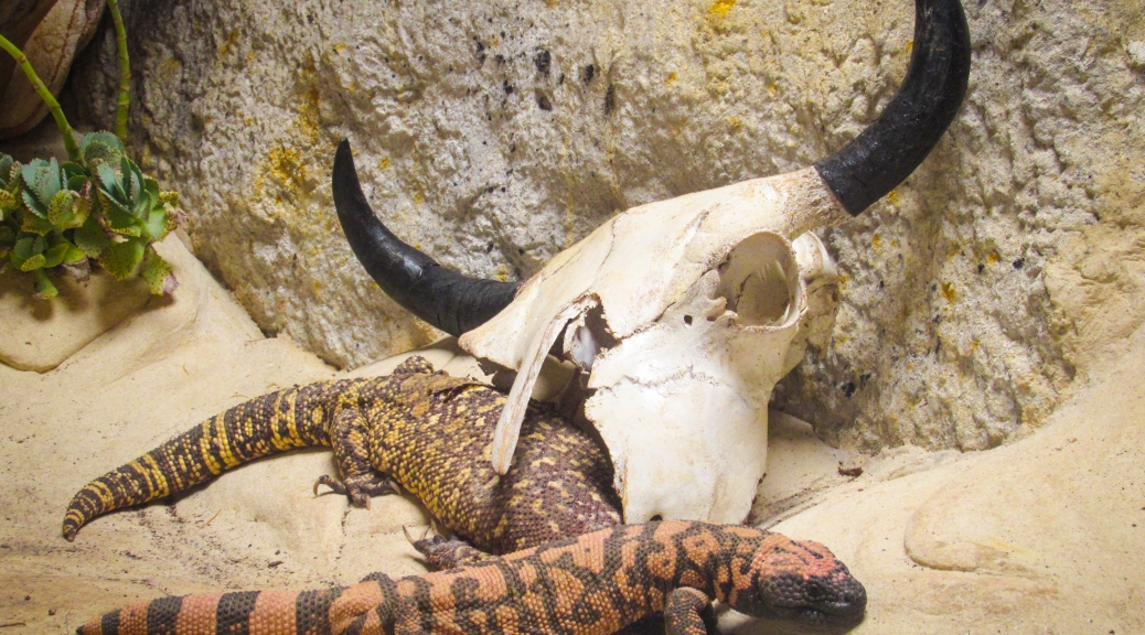
The most famous venomous lizards are the anguimorphs of the Helodermatidae family. From their discovery it was known that these lizards where venomous, as they present a pair of venomous glands in their lower jaws and various pairs of grooved teeth similar to those of venomous snakes with which they inject venom.

heloderma teethHelodermatid skull, in which we can see the sharp teeth with which they inject their venom. Image from Heloderma.net.

The helodermatis are carnivorous animals which feed on small mammals, birds, wall lizards, amphibians, invertebrates, eggs and carrion. Considering its generalist diet and that their prey are pretty defenceless, it is thought that venom evolved in these reptiles as a predator deterrent method, not as a hunting strategy.

2415413851_3d441fea6d_oPhoto by Walknboston of a Gila monster (Heloderma suspectum), in which we can see its black and yellow coloration, with which it warns its predators about its toxicity (aposematic coloration).

The Gila monster and the beaded lizard (Heloderma horridum) are slow animals which aren’t really dangerous to human beings. Yet their raising popularity as exotic pets has ended with some bite cases. The bite of a Gila monster causes some serious and burning pain, local edema, weakness, dizziness and nausea. Even if heavy bleeding is usually associated with bites, this isn’t due to some sort of anticoagulant substance but to the helodermatid’s sharp teeth and to the fact that to inject the venom they must chew their aggressor strongly , causing deep lacerations.

THE BEARDED DRAGON

The saurians of the genus Pogona are iguanians of the Agamidae family. These Australian reptiles are known as bearded dragons for the spines that they present on their throats. Even though they are adapted to live in arid places, the environmental temperature can affect the sex of their offspring.

Eastern_Bearded_Dragon_(Pogona_barbata)_(8243678492)Photo of an eastern bearded dragon in which we can see its yellow coloured mouth. Could it be that this coloration is indicating anything? Photo by Matt.

Bearded dragons are inoffensive animals, but there’s one species with a secret weapon. The eastern bearded dragon (Pogona barbata) is a venomous lizard but, while the rest of venomous reptiles only have one pair of venomous glands, the eastern bearded dragon has two pairs: two in its upper jaw and two in its lower jaw.

nature04328-f2.2Transversal section of the mouth of an eastern bearded dragon, in which we can see the incipient venomous glands both in its upper jaw (mxivg) and its lower jaw (mnivg). Image extracted from Fry, Vidal et al.

The venom they produce isn’t really strong (in human beings it only causes a minor swelling) and the glands are considered vestigial. Yet, the Toxicofera theory argues that the glands of the bearded dragon show us the primitive form which the first toxicoferan reptile would have presented, with two pairs of venom glands instead of a single pair like most current venomous reptiles.

THE BIG MONITORS

Everyone has heard about monitor lizards (anguimorphs of the Varanidae family). There are hundreds of documentaries about the Komodo dragon in which we are told that these animals have so many bacteria in their mouths that their bites inflict an infection, deadly enough to kill an adult bull. Yet recent studies have shown that the monitor’s poor buccal hygiene is not what causes the death of their victims.

Sans nom-35Perente or perentie (Varanus giganteus) a typical varanid, with long neck, strong legs, active metabolism and developed senses. Photo by Bernard Dupont.

Even if there are three frugivorous species, the rest are obligate carnivores. It has always been said that the mouth’s bacteria of the monitors is what causes the death of their prey, even if there isn’t any studies which prove it. In fact, in many studies it has been seen that the monitor’s saliva isn’t very different from that of other herbivorous reptiles.

3215319924_2fe90e244f_oPhoto in which we see the feared monitor’s saliva, specifically from an Asian water  monitor (Varanus salvator). Image by Lip Kee.

In a study, it was demonstrated that various species of monitor lizards present venom glands in their lower jaws. These glands are among the most complex venomous glands known of all reptiles. In the case of the Komodo dragon, these are compound glands with a larger posterior compartment and five smaller anterior compartments. These compartments have ducts that carry the venom between the teeth.

Even if varanids are closely related to snakes (they share, for example, a bifid tongue), these don’t present the snakes’ characteristic grooves in their teeth. This is due to the fact that instead of injecting the venom directly, monitor lizards use their serrated teeth to open a deep wound in their prey, through which the venom will enter the organism.

Varanus_priscus_skullSkull of megalania (Varanus priscus) in which we can see the teeth without gooves. This extinct monitor with more than 5 metres long, was the largest venomous animal known. Photo by Steven G. Johnson.

The utility of the venom for the predatory monitors is also supported by the large quantities of venom that they produce. In constrictor snakes that don’t utilise venom, the genes which codify the synthesis of venom are atrophied because of the great amount of energy required to produce it. Monitors, instead, secrete lots of venom with the slightest stimulation of their glands. This venom contains anticoagulant compounds which prevent the wound to close and also produces a cardiovascular shock in the animal by lowering the blood pressure.

Dragon_feedingA group of Komodo dragons (Varanus komodoensis) feeding on a recently killed pig. Image extracted from Bull, Jessop et al.

Even if we still don’t know for sure if the common ancestor of all these animals was venomous, nor if venom appeared independently in the different families, the relationship between the different members of the clade Toxicofera has been supported by posterior phylogenetic analyses. What we know is that venom is an extremely powerful weapon in the struggle for survival and that, even if snakes are the most numerous venomous reptiles, many other squamate species have been benefiting from the use of toxins, both for self-defence and to subjugate their prey.

REFERENCES

The following sources have been used during the elaboration of this entry:

Difusió-anglès

Iberian ophidians: nice snakes and venomous vipers

In my first blog entry I talked about the different kinds of snake that exist based on their dentition. In this entry, I’ll explain what species of ophidian we can find in the Iberian Peninsula, which species are venomous and which aren’t, and how we can identify the different species we can find when we are on the field. As we will see in this entry, snakes have been unfairly demonized, as the species in the Iberian Peninsula pose no threat to us.

INTRODUCTION

In the Iberian Peninsula we can find 13 different species of snakes, with representatives of three of the four types of dentition I talked about in my last post. There aren’t any proteroglyphous snake because the members of the Elapidae family are restricted to tropical and subtropical habitats. Most of the iberian species are snakes of the Colubridae family (aglyphous or opisthoglyphous) or vipers and adders of the Viperidae family (solenoglyphous).

Natrix maura bo
Viperine water snake (Natrix maura), aglyphous
Malpolon bo
Montpellier snake (Malpolon monspessulanus), opisthoglyphous
Vipera latastei bo
Snub-nosed viper (Vipera latastei), solenoglyphous

COLUBRIDS vs. VIPERS

When we find a snake in the wild it’s important to know if that animal is a colubrid or a viper. Bites from Iberian colubrids are mostly harmless because they have either an unspecialized non-venomous dentition (aglyphous) or posterior venomous fangs (opisthoglyphous) which usually doesn’t inject venom and even if they do, normally they don’t inject enough venom for it to be dangerous. On the other hand, as Iberian vipers are solenoglyphous, they inject large quantities of venom, being vipers responsible for most of the snake bite accidents in Spain. Yet, bites are extremely rare, and most happen after a too prolonged manipulation of the animal.

To identify a snake as a colubrid or a viper there are some anatomical characteristics which tell them apart. These characteristics are usually only applicable to iberian ophidians; species from outside the Iberian Peninsula may present different combinations of characters.

The most cited character is the pupil. Normally vipers show an elliptic, slit-like pupil, while colubrids present a round pupil. However, this character is variable, because with low-light conditions a viper’s pupil may look round as the eyes of these animals can adapt to darkness.

PUPILA
Colubrid with round pupil (ringed snake, Natrix natrix) and viperid with elliptic pupil (snub-nosed viper, Vipera latastei). Photos by Honorio Iglesias.

The second character refers to the shape of the body. While colubrids are mostly thin, have an undifferentiated neck and a long slim tail, vipers have a triangular-shaped head with a neck differentiated from the body, and a short and conic tail.

BODYYY
Aesculapian snake (Zamenis longissimus) and Baskian viper (Vipera seoanei, photo by Daniel Gómez)

Although it may be difficult to look at, scales can be useful to tell colubrids and vipers apart. Vipers always present keeled scales, which have a little keel-like protuberance longitudinally on it. On the other side, even though they can have some keeled scales, most colubrids present smooth scales.

SCALES
Smooth scales of a horseshoe whip snake (Hemorrhois hippocrepis, photo by Saúl Yubero) and keeled scales of an asp viper (Vipera aspis, photo by Grégoire Meier)

Finally, while colubrids are active animals and usually flee before we can get close to them, vipers rely on their camouflage to avoid predation. Therefore, they stay still so we can’t see them, and may bite if they feel cornered.

IBERIAN OPHIDIANS

Colubridae family:

Coronella genus: Known as smooth snakes. In the Iberian Peninsula we can find the northern smooth snake (Coronella austriaca) which presents a dark mask-like spot covering from the nasal openings up to the neck and dark irregular markings on its back, and the southern smooth snake (Coronella girondica) which presents a pair of parietal marks and dark transversal spots on its back.

Coronella aust gir
Northern smooth snake (Coronella austriaca, left, photo by Christian Fischer) and southern smooth snake (Coronella girondica, right, photo by Evaristo Corral)

Hierophis genus: The green whip snake or western whip snake (Hierophis viridiflavus) is a brightly-coloured snake with a pattern of black, yellow and green spots over its body. Even though they can grow up to 170 cm of length they are not venomous. It can be usually found from temperate forests to crop fields, and even in abandoned buildings.

Hierophis viri
Green whip snake (Hierophis viridiflavus), juvenile (left, by Polypterus) and adult (right)

Natrix genus: Commonly known as water snakes due to their affinity for aquatic habitats. In the Iberian Peninsula we can find two species, the viperine water snake (Natrix maura) named after its zigzag marking and its keeled scales similar to a viper, and the grass or iberian ringed snake (Natrix astreptophora) which presents reddish pupils, an extremely variable coloration and a black “ring” in juvenile individuals.

Natrix mau nat
Viperine water snake (Natrix maura, left, photo by Honorio Iglesias) and iberian ringed snake (Natrix astreptophora, right photo of Fafner).

Zamenis genus: The Aesculapian snake (Zamenis longissimus) is a slim, long and harmless colubrid with a characteristically narrow and elongated skull. It is normally found on forested areas, with different microclimatic variations to aid it on its thermoregulation. This species is the one represented coiled around the rod of Aesculapius and the Bowl of Hygieia, symbols of medicine and pharmacy respectively.

Zamensis long
Aesculapian snake (Zamenis longissimus) (left by Amiralles).

Hemorrhois genus: The horseshoe whip snake (Hemorrhois hippocrepis) is an aglyphous colubrid that, even if it may bite if touched or grabbed, it’s not considered a venomous species. It presents a transversal mark on its head from one eye to the other, and another mark in the shape of a horseshoe on its neck, which gives this species its common name. It’s a species typical of rocky habitats.

Hemorrhois hippo
Horseshoe whip snake (Hemorrhois hippocrepis). Photos by Accipiter and Raúl León.

Rhinechis genus: The ladder snake (Rhinechis scalaris) receives its common name due to the stripes that juvenile specimens present on their back, similar to a ladder, even though adult individuals may present only longitudinal stripes on their body without any transversal marks connecting them. Despite being an apparently aggressive snake, it rarely bites and is harmless to human beings.

Rhinechis sca
Ladder snake (Rhinechis scalaris). Photos by Matt Wilson (left) and by Fernando Fañanás (right).

Macroprotodon genus: This is one of the few venomous species in the Peninsula. The western false smooth snake (Macroprotodon brevis) is an animal common on many different Mediterranean habitats. Even if it’s venomous, its small opisthogyphous mouth and its calm behavior make it totally harmless. It is characterised by a dark mark on the back of its head, and its short and flattened skull.

Macroprotodon brev
Western false smooth snake (Macroprotodon brevis). Photos by Saúl Yubero and Amiralles, respectively.

Malpolon genus: With specimens growing up to two and a half meters of length, the Montpellier snake (Malpolon monspessulanus) is the largest ophidian of the peninsula. Due to its opisthoglyphous dentition it normally doesn’t inject venom when biting (which is extremely rare), but larger individuals with much wider mouths may inject venom, but to cause symptoms it should hold its bite for a long period of time (most bites, even if rare, are dry warning bites). It is easily recognisable for its prominent eyebrows which give it a ferocious look.

Malpolon mons
Montpellier snake (Malpolon monspessulanus). Photos by Herpetofauna and RuizAraFoto respectively.

RuizAraFoto

Viperidae family:

There’s only one genus of vipers on the Iberian Peninsula with three representative species. Vipers and adders usually have a wide and triangular head, a lightly elevated snout and usually present a zigzag pattern on their back which help them camouflage. The three Iberian species are venomous, but thanks to modern medicine, their ocasional bites aren’t harmful to human beings. The asp viper (Vipera aspis), the most venomous snake in the peninsula, presents grey, golden or yellow scales, with black or green spots. The snub-nosed viper (Vipera latastei) is the most common viper in the peninsula and its coloration varies from brown to grey. Finally the Baskian or Portuguese viper (Vipera seoanei) is a middle-sized viper and with a highly polymorphic pattern.

Vipera asp lat seo
Asp viper (Vipera aspis, top left, photo by Felix Reimann), snub-nosed viper (Vipera latastei, top right, photo by Honorio Iglesias) and Baskian viper (Vipera seoanei, bottom, photo by Andre Schmid).

As we have seen, snakes and vipers aren’t as bad as they are portrayed to be. Most species flee from human beings, and accidents and bites happen when we force them to interact with us too much. Also, ophidians help farmers and agriculturers by hunting and eating species traditionally seen as vermin. If we leave snakes and vipers alone, we will be able to enjoy the beauty of this animals in peace.

REFERENCES

The following sources have been consulted in the elaboration of this entry:

If you enjoyed this article, please share it on social networks tospread it. The aim of the blog, after all, is to spread science and reachas many people as possible.

Licencia Creative Commons
Licencia Creative Commons Atribución-NoComercial-CompartirIgual 4.0 Internacional.