Arxiu d'etiquetes: lizard

Desert reptiles

Deserts are some of the most extreme habitats on the planet. The Sahara, the Gobi and the Sonora are some examples of warm deserts where the high temperatures and the lack of water pose a great challenge to animals that live in them. Reptiles are one of the animal groups that present the most incredible adaptations for life in deserts. In this entry we’ll explain the difficulties that desert reptiles must face in order to survive, and we’ll introduce you to different species of snakes and lizards that in the deserts have found their home.

REPTILES IN THE DESERT

The characteristic which unites all deserts is the scarce precipitation as, unlike most people think, not all deserts present high temperatures (there are also cold deserts, like the Arctic and the Antarctic, both in danger because of the climate change). Reptiles thrive better in warm deserts than in cold deserts, because the low temperatures would not allow them to develop their life activity.

aavikko
Map by Vzb83 of the warm deserts, both arid and semiarid, of the world.

Warm deserts not always have extremely high temperatures. While during the day temperatures may rise up to 45°C, when the sun goes down temperatures fall below freezing point, creating daily oscillations of up to 22°C. The different desert reptiles, being poikilotherms and ectotherms, use different behavioural strategies in order to avoid overheating during the day and to keep their body heat during the night (for example, climbing to elevated areas or sleeping in burrows).

The Namaqua chameleon (Chamaleo namaquensis) regulates its body heat changing its colouration. During sunrise it is black in colour in order to absorb as much radiation of the sun and activate its metabolism. When temperatures become higher, it turns white to reflect solar radiation. Video from BBC.

As we have already stated, the main characteristic of any desert is the lack of water. Generally, in a desert, it rains less than 250 mm of water a year. The scaly and impervious skin of reptiles prevents the loss of water, and their faeces contain uric acid which, compared to urea, is much less soluble in water, allowing them to retain more liquids. Most desert reptiles extract the water they need from their food and some drink water from the dew.

Both the extreme temperatures and the shortage of precipitations make the desert a place with very few living beings. Vegetation is scarce and animals are usually small and secretive. This lack of resources causes desert reptiles to be usually smaller than their cousins from more benevolent environments. Also, these saurians usually exploit any available food resource, although they think twice before wasting their precious energy to get their next meal.

SAND SNAKES

In many sandy deserts we can find various species of snakes (and legless lizards) that have adapted to a life among the dunes. Many of these ophidians share a locomotion method called “sidewinding”, in which they raise their head and neck from the ground and move them laterally while the rest of the body stays on the ground. When they place their head on the ground again they raise their body, making these snakes move laterally in a 45° angle. This method of locomotion makes these snakes move more efficiently in an unstable terrain. It also reduces the contact of their body with an extremely hot substrate, as the body of these ophidians only touches the ground in two points at a time.

As we can see in this video from RoyalPanthera, sidewinding allows desert snakes to move minimizing the contact with the hot terrain.

Many desert ophidians bury themselves in the sand both to avoid sun exposure and to blend in and catch their prey unaware. This has made many desert-dwelling snakes very sensitive to vibrations generated by their prey as it moves through the sand. In addition some species present an overly developed rostral scale (the scale at the tip of their snout), being much thicker in order to aid during excavation in sandy soils.

heterodon_nasicus2
An example of this are the North American snakes of the Heterodon genus, also known as hog-nosed snakes, as they present an elevated rostral scale giving their snout a characteristic shape. Photo of Heterodon nasicus by Dawson.

The horned vipers of the Cerastes genus also present various characteristics that facilitate life in the deserts. These vipers evade high temperatures becoming active at night and they spend the day buried in the sand. Their hunting method consists in burying themselves waiting for a prey to pass by, this way saving most of their energy. It is believed that their horn-shaped supraocular scales cover their eyes when they are buried in order to protect them from the sand.

10680524213_5584c4ddb8_o
Photo by Tambako The Jaguar of a Sahara sand viper (Cerastes vipera), a species from North Africa and the Sinai Peninsula.

SPINY CRITTERS

In different deserts of the world we find reptiles with their bodies covered in spines. This not only provides them with certain protection against predators, but is also helps them blend in in a habitat with plenty of thorny plants. Two of these animals are members of the Iguania suborder: the thorny devil and the horned lizards.

thorny_-_christopher_watson
Photo of a thorny devil (Moloch horridus) by Christopher Watson.

The thorny devil (Moloch horridus) is an agamid that lives in the Australian sandy deserts. This lizard presents spines all over its body, making it difficult for its predators to swallow. It also has a protuberance behind its head that acts as a fat storage.  When it feels threatened, it hides its real head between its legs and it exposes its neck protuberance as a decoy head. Probably, the most interesting adaptation of this animal is the system of small grooves among its scales, which collect any water that contacts its skin and conducts it directly to its mouth.

Horned lizards (Phrynosoma genus, affectionately called “horny toads”) are iguanids which are found in different arid habitats of North America. Similarly to the thorny devil, their body is covered in spines making them hard to eat for their predators. Also, when they are caught, they inflate their bodies to make the task even more difficult. Finally, some species like the Texas horned lizard (Phrynosoma cornutum) are known for their autohaemorrhagic abilities: when they feel cornered they squirt a stream of stinky blood from their eyes which scares away most predators.

federal_horned_toad_pic_crop
Photo from the U.S. Fish & Wildlife Service of a Texan horned lizard (Phrynosoma cornutum).

As you have seen, in the deserts we can find reptiles with some of the most inventive (and disturbing) adaptations of the world. These are only a few examples of the astonishing diversity of squamates that are found in the deserts of the world, which only seek to survive the harsh conditions of these extreme environments. Sometimes, it’s just a matter to avoid burning your feet with the hot sand.

Video from BBCWorldwide of a shovel snouted lizard (Zeros anchietae) making the “thermal dance” in order to diminish the contact with the hot sand.

REFERENCES

The following sources have been consulted during the elaboration of this entry:

difusio-angles

Monsters and dragons: Venomous lizards

When we think about venomous animals most people think about the same ones. Usually, we think about spiders, scorpions and snakes, despite knowing there are also venomous amphibians, fishes and mammals. Even if snakes are the best known venomous reptiles, in time we have learned that they are not the only group that present venomous glands and that many other reptiles also have the capacity of injecting venom. In this entry we’ll get to know the least known venomous saurians and we’ll try to explain their relationship with snakes.

EVOLUTION OF VENOM IN REPTILES

Everybody is familiar with the toxic abilities of snakes. Traditionally it was believed that venom evolved independently in the different groups of venomous snakes (colubrids, elapids and viperids) and in a lizard family (the helodermatids). Yet this vision has changed over the years and with the discovery of other species of venomous squamates.

Venom_extraction
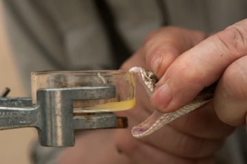
The venom of many animals is used for both antivenom development and pharmacological research of analgesics and other medicines. Photo of the extraction of venom from a saw-scaled viper (Echis carinatus), by Kalyan Varma (Image under a GNU license).

Currently, it’s been shown that there are different species of saurian which present glands and organs capable of injecting venom, along with many other species with genetic material related to venom production (even if most aren’t venomous). This occurs, for example, in many apparently non-venomous snakes and lizards that retain genetic material related to the synthesis of venom. This has caused many scientists to group these reptiles under a common clade called Toxicofera, “those who bear toxins”.

This new clade includes the different squamosal taxa, which are believed to have had a venomous common ancestor. These groups are:

  • Ophidia: Ophidians, snakes.
Indian_wolf_snake_(Lycodon_aulicus)_Photograph_By_Shantanu_Kuveskar
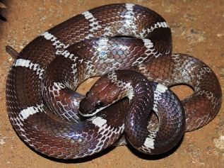
Indian wolf snake (Lycodon aulicus), example of an ophidian. Photo by Shantanu Kuveskar.
  • Iguania: Iguanas, agamas and chameleons.
6968443212_4b3f4fbd7f_o
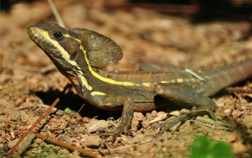
Brown basilisk (Basiliscus vittatus), example of an iguanian. Photo by Steve Harbula.
Real_Lanthanotus_borneensisEarless monitor lizard (Lanthanotus borneensis), example of an anguimorph. Photo by Kulbelbolka.

Even though most current iguanians and anguimorphs don’t present venom, the Toxicofera theory proposes that many species would have lost their capacity to inject venom secondarily. Below we’ll present some of the lesser known venomous saurians.

MONSTERS OF THE NEW WORLD

The most famous venomous lizards are the anguimorphs of the Helodermatidae family. From their discovery it was known that these lizards where venomous, as they present a pair of venomous glands in their lower jaws and various pairs of grooved teeth similar to those of venomous snakes with which they inject venom.

heloderma teeth
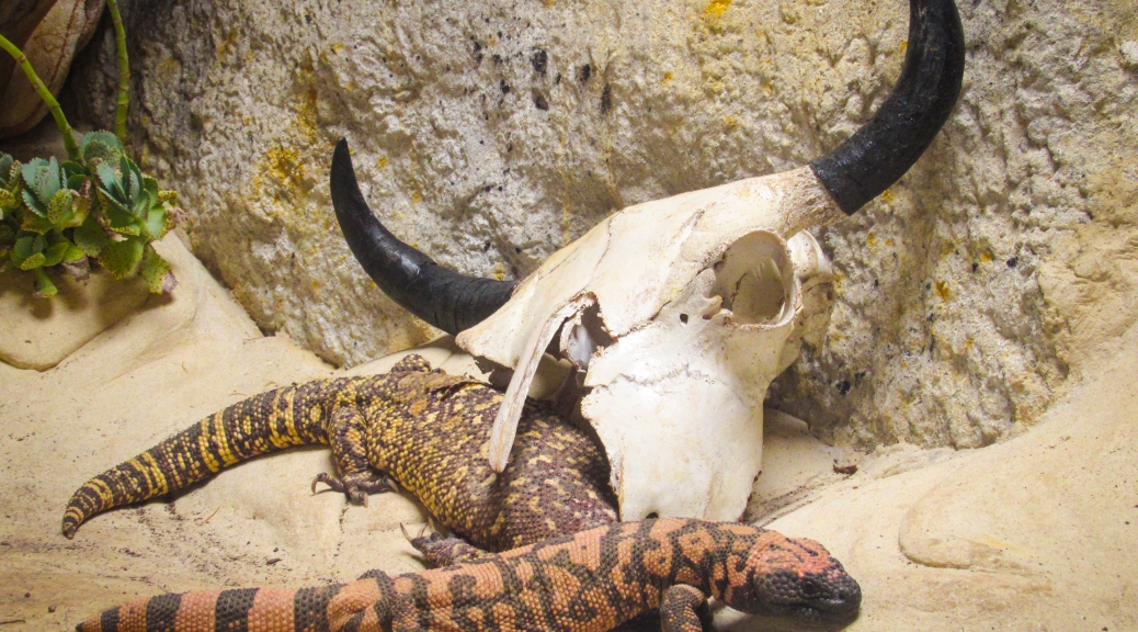
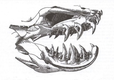
Helodermatid skull, in which we can see the sharp teeth with which they inject their venom. Image from Heloderma.net.

The helodermatis are carnivorous animals which feed on small mammals, birds, wall lizards, amphibians, invertebrates, eggs and carrion. Considering its generalist diet and that their prey are pretty defenceless, it is thought that venom evolved in these reptiles as a predator deterrent method, not as a hunting strategy.

2415413851_3d441fea6d_oPhoto by Walknboston of a Gila monster (Heloderma suspectum), in which we can see its black and yellow coloration, with which it warns its predators about its toxicity (aposematic coloration).

The Gila monster and the beaded lizard (Heloderma horridum) are slow animals which aren’t really dangerous to human beings. Yet their raising popularity as exotic pets has ended with some bite cases. The bite of a Gila monster causes some serious and burning pain, local edema, weakness, dizziness and nausea. Even if heavy bleeding is usually associated with bites, this isn’t due to some sort of anticoagulant substance but to the helodermatid’s sharp teeth and to the fact that to inject the venom they must chew their aggressor strongly , causing deep lacerations.

THE BEARDED DRAGON

The saurians of the genus Pogona are iguanians of the Agamidae family. These Australian reptiles are known as bearded dragons for the spines that they present on their throats. Even though they are adapted to live in arid places, the environmental temperature can affect the sex of their offspring.

Eastern_Bearded_Dragon_(Pogona_barbata)_(8243678492)
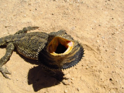
Photo of an eastern bearded dragon in which we can see its yellow coloured mouth. Could it be that this coloration is indicating anything? Photo by Matt.

Bearded dragons are inoffensive animals, but there’s one species with a secret weapon. The eastern bearded dragon (Pogona barbata) is a venomous lizard but, while the rest of venomous reptiles only have one pair of venomous glands, the eastern bearded dragon has two pairs: two in its upper jaw and two in its lower jaw.

nature04328-f2.2
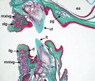
Transversal section of the mouth of an eastern bearded dragon, in which we can see the incipient venomous glands both in its upper jaw (mxivg) and its lower jaw (mnivg). Image extracted from Fry, Vidal et al.

The venom they produce isn’t really strong (in human beings it only causes a minor swelling) and the glands are considered vestigial. Yet, the Toxicofera theory argues that the glands of the bearded dragon show us the primitive form which the first toxicoferan reptile would have presented, with two pairs of venom glands instead of a single pair like most current venomous reptiles.

THE BIG MONITORS

Everyone has heard about monitor lizards (anguimorphs of the Varanidae family). There are hundreds of documentaries about the Komodo dragon in which we are told that these animals have so many bacteria in their mouths that their bites inflict an infection, deadly enough to kill an adult bull. Yet recent studies have shown that the monitor’s poor buccal hygiene is not what causes the death of their victims.

Sans nom-35
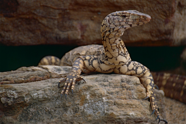
Perente or perentie (Varanus giganteus) a typical varanid, with long neck, strong legs, active metabolism and developed senses. Photo by Bernard Dupont.

Even if there are three frugivorous species, the rest are obligate carnivores. It has always been said that the mouth’s bacteria of the monitors is what causes the death of their prey, even if there isn’t any studies which prove it. In fact, in many studies it has been seen that the monitor’s saliva isn’t very different from that of other herbivorous reptiles.

3215319924_2fe90e244f_o
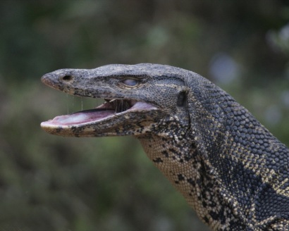
Photo in which we see the feared monitor’s saliva, specifically from an Asian water  monitor (Varanus salvator). Image by Lip Kee.

In a study, it was demonstrated that various species of monitor lizards present venom glands in their lower jaws. These glands are among the most complex venomous glands known of all reptiles. In the case of the Komodo dragon, these are compound glands with a larger posterior compartment and five smaller anterior compartments. These compartments have ducts that carry the venom between the teeth.

Even if varanids are closely related to snakes (they share, for example, a bifid tongue), these don’t present the snakes’ characteristic grooves in their teeth. This is due to the fact that instead of injecting the venom directly, monitor lizards use their serrated teeth to open a deep wound in their prey, through which the venom will enter the organism.

Varanus_priscus_skull
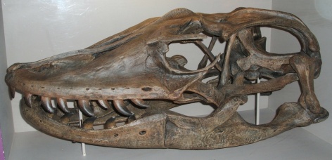
Skull of megalania (Varanus priscus) in which we can see the teeth without gooves. This extinct monitor with more than 5 metres long, was the largest venomous animal known. Photo by Steven G. Johnson.

The utility of the venom for the predatory monitors is also supported by the large quantities of venom that they produce. In constrictor snakes that don’t utilise venom, the genes which codify the synthesis of venom are atrophied because of the great amount of energy required to produce it. Monitors, instead, secrete lots of venom with the slightest stimulation of their glands. This venom contains anticoagulant compounds which prevent the wound to close and also produces a cardiovascular shock in the animal by lowering the blood pressure.

Dragon_feeding
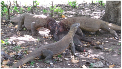
A group of Komodo dragons (Varanus komodoensis) feeding on a recently killed pig. Image extracted from Bull, Jessop et al.

Even if we still don’t know for sure if the common ancestor of all these animals was venomous, nor if venom appeared independently in the different families, the relationship between the different members of the clade Toxicofera has been supported by posterior phylogenetic analyses. What we know is that venom is an extremely powerful weapon in the struggle for survival and that, even if snakes are the most numerous venomous reptiles, many other squamate species have been benefiting from the use of toxins, both for self-defence and to subjugate their prey.

REFERENCES

The following sources have been used during the elaboration of this entry:

Difusió-anglès

Reptiles and mammals: same origin, different stories

Did mammals evolve from reptiles? The truth is they didn’t. Reptiles and mammals both have independent evolutionary histories that separated soon after the apparition of the so-called amniotic egg, which allowed the babies of these animals to be born outside of water. Previously, we talked about the origin of vertebrates and about how they managed to get out of the sea to start walking on land for the first time. In this entry we’ll explain how the ancestors of reptiles and mammals, the AMNIOTES, became independent of the aquatic medium and became the dominant land animals.

THE AMNIOTIC EGG

The characteristic that unites reptiles and mammals in the same group is the amniotic egg. While amphibian eggs are relatively small and only have one inner membrane, the eggs of amniotes are much bigger and present various membranes protecting the embryo and keeping it in an aqueous medium. The outer layer is the eggshell which, apart from offering physical protection to the embryo, prevents water loss and its porosity allows gas interchange. Beneath the eggshell we can find the next membranes:

512px-Crocodile_Egg_Diagram.svgDiagram of a crocodile egg: 1. eggshell 2. yolk sac 3. yolk (nutrients) 4. vessels 5. amnion 6. chorion 7. air 8. alantois 9. albumin (white of the egg) 10. amniotic sac 11. embryo 12. amniotic fluid. Image by Amelia P.
  • Chorion: The first inner membrane, which offers protection and, together with the amnion, forms the amniotic sac. Also, being in contact with the eggshell, it participates in gas interchange, bringing oxygen from the outside to the embryo and carbon dioxide from the embryo to the outside.
  • Amnion: Membrane that surrounds the embryo and constitutes a part of the amniotic sac. It offers an aqueous medium for the embryo and connects it with the yolk sac (a structure that brings food and that is also found in fish and amphibians).
  • Allantois: The third layer, it is used as a storage for nitrogen waste products, and together with the chorion, helps in gas interchange.
512px-Amphibian_Egg_Diagram.svgDiagram of an amphibian egg: 1. jelly capsule 2. vitelline membrane 3. perivitelline fluid 4. yolk 5. embryo. Image by Separe3g.

All these different kinds of membranes eliminate the need amphibians had of laying their eggs in water. Also, unlike amphibians, amniotes don’t go through a gilled larval stage, but are instead born as miniature adults, with lungs and legs (at least those that have them). All these made the first amniotes completely independent of the aquatic medium.

AMNIOTE ORIGINS

The first amniotes evolved around 312 million years ago from reptiliomorph tetrapods. At the end of the Carboniferous period lots of tropical forests where the great primitive amphibians lived disappeared, leaving a colder and drier climate. This ended with many of the big amphibians of that time, allowing the amniotes to occupy new habitats.

Solenodonsaurus1DBReconstruction of Solenodonsaurus janenschi, one of the candidates in being the first amniote, which lived around 320-305 million years ago in what is now the Czech Republic. Reconstruction by Dmitry Bogdanov.

CHARACTERISTICS

These early amniotes had a series of characteristics that set them apart from their semiaquatic ancestors:

  • Horny claws (amphibians don’t have claws) and keratinized skin that prevents water loss.
  • Bigger large intestine and higher density of renal tubules to increase water reabsorption.
  • Specialized lacrimal glands and a third membrane in the eye (nictitating membrane) which keep the eye wet.
  • Larger lungs.
  • Loss of the lateral line (sensory organ present in fish and amphibians).

The skeleton and musculature also evolved offering better mobility and agility on a terrestrial medium. The first amniotes presented ribs that encircled their body converging at the sternum, making their inner organs more secure, and a series of muscular receptors offered them better agility and coordination during locomotion.

AMNIOTE SKULLS

Traditionally, the different amniotes were classified based on the structure of their cranium. The characteristic used to classify them was the presence of temporal openings (fenestrae), by which we have three groups:

  • Anapsids (“no arches”): No temporal openings (turtles).
Skull_anapsida_1Diagram of an anapsid skull, by Preto(m).
  • Synapsids (“fused arches”): With only one temporal opening (mammals).
Skull_synapsida_1Diagram of a synapsid skull, by Preto(m).
  • Diapsids (“two arches”): With two temporal openings (reptiles, including birds).
Skull_diapsida_1Diagram of a diapsid skull, by Preto(m).

Previously it was believed that the first amniotes presented an anapsid skull (without openings, like turtles) and that subsequently they separated into synapsids and diapsids (the temporal openings formed “arches” that offered new anchor points for the jaw’s musculature). Yet, it has been discovered that this three-group classification is not valid.

Even though we still believe that the first amniotes were anapsid, it is currently known that these, soon after their apparition, separated into two different lineages: the synapsids (clade Synapsida) and the sauropsids (clade Sauropsida).

SYNAPSIDA

This lineage includes mammals and their amniote ancestors. Even though the first synapsids like Archaeothyris looked externally like lizards, they were more closely related to mammals, as they shared one temporal fenestrae where the jaw muscles passed through.

Archaeothyris.svgDrawing of the skull of Archaeothyris, which is thougth to be one of the first synapsids that lived around 306 million years ago in Nova Scotia. Drawing by Gretarsson.

The ancestors of mammals were previously known as “mammal-like reptiles”, as it was thought that mammals had evolved from primitive reptiles. Currently it’s accepted that synapsids form a different lineage independent of reptiles, and that they share a series of evolutionary trends that makes them closer to modern mammals: the apparition of different kinds of teeth, a mandible made of one single bone, the vertical posture of their limbs, etc…

Dimetrodon_grandisReconstruction of Dimetrodon grandis, one of the better known synapsids, from about 280 million years ago. Reconstruction by Dmitry Bogdanov.

Even though most modern mammals don’t lay eggs and give birth to live offspring, all groups maintain the amniote’s three characteristic membranes (amnion, chorion and allantois) during embryonic development.

SAUROPSIDA

Sauropsids include current reptiles and their amniote ancestors. Currently, in many scientific papers the word “sauropsid” is used instead of “reptile” when discussing phylogenies, as the sauropsids also includes birds. The first sauropsids were probably anapsids, and soon after their appearance they separated into two groups: the Parareptilia which conserved anapsid skull, and the Eureptilia which include the diapsids (current reptiles and birds).

Traditional_ReptiliaEvolutionary tree of current vertebrates, in which green color marks the groups previously included inside reptiles. As you can see, the traditional conception of "reptile" includes the ancestors of mammals and excludes birds. Image by Petter Bøckman.

Diapsids are currently the most diversified group of land vertebrates. They diversified greatly in the late Permian period (about 254 million years ago), just before the Mesozoic (the Age of Reptiles). These can be divided into two main groups: the Lepidsaurs and the Archosaurs, both with representatives in our days.

LEPIDOSAURIA: SMALL AND PLENTIFUL

Lepidosaurs (literally “reptiles with scales”) appeared in the early Triassic (around 247 million years ago) and, even if most of them didn’t grow to big sizes, they are currently the largest group of non-avian reptiles. These are characterized by presenting a transversal cloacal slit, by having overlapping scales and shedding their skin whole or in patches and by other skeletal characters.

Rat_Snake_moulted_skinShed skin of a rat snake. Photo by Mylittlefinger.

The current lepidosaurs belong to one of two different orders:

  • Order Rhynchocephalia: That includes the two species of tuatara. Currently endangered, they are considered living fossils because they present skulls and characteristics similar to the Mesozoic diapsids.
Sphenodon_punctatus_(5)Photo of a tuatara (Sphenodon punctatus), by Tim Vickers.
  • Order Squamata: Current squamates include iguanas, chameleons, geckoes, skinks, snakes and other legless lizards. With more than 9000 living species, squamates are a large group with a wide array of adaptations and survival strategies.
Sin títuloPhotos of some squamates, from left to right and from top to bottom: Green iguana (Iguana iguana, by Cary Bass), king cobra (Ophiophaga Hannah, by Michael Allen Smith), Mexican mole lizard (Bipes biporus, by Marlin Harms) and Indian chameleon (Chamaeleo zeylanicus, by Shantanu Kuveskar).

ARCHOSAURIA: ANCIENT KINGS

Archosaurs (literally “ruling reptiles”) were the dominant group of land animals during the Mesozoic. These conquered all possible habitats until the extinction of most groups at the end of the Cretaceous period. Some of the extinct groups were the pseudosuchians (relatives of modern crocodiles, order Crocodylia), the pterosaurs (large flying reptiles) and the dinosaurs (excepting birds, clade Aves).

Massospondylus_Skull_Steveoc_86Drawing of the skull of the dinosaur Massospondylus in which we can see the different characteristic openings of diapsid archosaurs. Image by Steveoc 86.

As you see, both groups of modern archosaurs couldn’t be more different. Yet, crocodiles and birds share a common ancestor, and they are both more closely related with each other than with the rest of reptiles.

Yellow-billed_stork_kazingaPhoto of two species of modern arcosaurs: a Nile crocodile (Crocodylus niloticus) and a yellow-billed stork (Mycteria ibis). Photo by Tom Tarrant.

AND WHAT ABOUT TURTLES?

Turtles (order Testudines) have always been a group difficult to classify. Turtles are the only living amniotes with an anapsid skull, without any post-ocular opening. That’s why previously they had been classified as descendants of primitive amniotes (clade Anapsida, currently disused) or as primitive anapsid sauropsids (inside the Parareptilia clade)

KONICA MINOLTA DIGITAL CAMERASkeleton of the extinct tortoise Meiolania platyceps which lived in New Caledonia until 3000 years ago. In this photo it can be seen the compact cranium without openings. Photo by Fanny Schertzer.

Recent molecular studies have revealed that turtles are actually diapsids that lost their temporal openings secondarily. What still divides the scientific community is if testudines are more closely related to Lepidosauromorphs (lepidosaurs and their ancestors) or to Archosauromorphs (archosaurs and their ancestors).

Leopard_tortoiseIndividual leopard tortoise (Stigmochelys pardalis) from Tanzania. Photo by Charles J. Sharp.

As you have seen, the evolution of amniotes is an extremely complex matter. We hope that with this entry some concepts have been clarified:

  1. Mammals (synapsids) come from an evolutionary lineage different from that of reptiles (sauropsids).
  2. Sauropsids include traditional reptiles (lepidosaurs, archosaurs and turtes) and birds (inside archosaurs).
  3. There’s still so much to investigate about the placement of turtles (testudines) in the evolutionary tree of sauropsids.
Figure_29_04_03Modified diagram about the evolutionary relationships of the different amniote groups.

REFERENCES

During the elaboration of this entry the following sources have been consulted:

Difusió-anglès

Limb regeneration, from the axolotl to human beings

The regeneration of lost or damaged body parts in animals is known from many centuries ago. In 1740 the naturalist Abraham Trembley observed a small cnidarian that could regenerate its head if it was cut off, so he called it Hydra, in reference to the monster from Greek mythology that could grow back its multiple heads if they were cut off. Afterwards, it was discovered that there were many other species of animals with regenerative abilities. In this entry we’ll talk about these animals.

Regeneration in the animal kingdom

Regeneration of body parts is more widespread between the different groups of invertebrates than it is between the vertebrates. This process can be bidirectional, in which both parts of the animal regenerate their missing parts to form two animals (just like the hydra, planarians, earthworms and starfishes) or unidirectional, in which the animal loses an extremity but it just regenerates, without forming two animals (arthropods, molluscs and vertebrates). In vertebrates, fishes and amphibians are the ones that present the greatest regenerative capacities, although many lizards and some mammals are able to regrow their tails.

ch14f01Image by Matthew McClements about bidirectional regeneration in planarians, hydras and seastars. Extracted from Wolbert's Principles of Development.

Regeneration can be done by two different ways:

  • Regeneration without active cellular proliferation or “morphallaxis”. In this type, the absent body part is regrown through remodelling of pre-existing cells. This is what happens in the Hydra, in which lost body parts are regenerated without the creation of new material. So, if a hydra is cut in half, we’ll obtain two smaller versions of the original hydra.
Video about an experiment in which an Hydra has been cut in different pieces. Video by Apnea.
  • Regeneration with cellular proliferation or “epimorphosis”. In this type, the lost part is regenerated via cellular proliferation, it is “newly created”. In most cases, it happens through the formation of a specialized structure called blastema, a mass of undifferentiated cells which appears during phenomena of cellular regeneration.

Almost all groups of animals with regenerative capacities present regeneration with blastema formation. Yet the origin of the blastemal stem cells varies between groups. While planarians present pluripotent (that can differentiate to any kind of cell type) stem cells all along their bodies, vertebrates have specific cells in each type of tissue (cartilage, muscle, skin…) that only regenerate cells of the tissue they come from.

In land vertebrates, lizards and urodeles are the ones that present the most powerful regenerative abilities. Down below we’ll see how they regenerate and the applications it has in modern human medicine.

Expendable tails

When you are a small animal that is being chased by a cat or any other predator, it probably is better for you to lose your precious tail than to lose your life. Some terrestrial vertebrates have evolved following this philosophy, and they are able to shed off their tails voluntarily through a process called caudal autotomy. This allows them to escape from their predators, which are entertained with the still moving lost tail.

 Video in which we can see how some lizards like this red-tailed vanzosaur (Vanzosaura rubricauda) have brightly coloured tails to attract the attention of predators. Video by Jonnytropics.

Autotomy or self-amputation, is defined as a behaviour in which the animal can shed off one or more body parts. Caudal autotomy is found in many species of reptiles and in two species of spiny mouse of the genus Acomys. In reptiles we can find caudal autotomy in lacertids, geckos, skinks and tuataras.

Acomys.cahirinus.cahirinus.6872Foto of a Cairo spiny mouse (Acomys cahirinus), a mammal which is able to shed and regrow its tail. Photo by Olaf Leillinger.

In reptiles, the fracture of the tail happens in specific areas of the caudal vertebras which are naturally weakened. The autotomy may happen in two different ways: intravertebral autotomy, in which the vertebra at the centre of the tail have transversal fracture planes prepared to break if they are pressed hard enough, and intervertebral autotomy, where the tail breaks between vertebras by muscular constriction.

0001-3765-aabc-201520130298-gf03Tridimensional model of the fracture planes on the tail of a lizard and the regeneration post-autotomy of a cartilaginous tube. Image extracted from Joana D. C. G. de Amorim et al.

Caudal autotomy allows the animal to escape, but it isn’t without cost. Many reptiles use their tails as a reserve of fat and losing this energy store is usually detrimental for the animal. That’s why many lizards, once the threat has disappeared, look for their lost tail and eat it, to at least regain the energy it had as fat. In addition, regenerating a new tail requires a great expenditure of energy.

DSCN9467Photo of a Catalonian wall lizard (Podarcis liolepis) that has shed its tail. Photo by David López Bosch.

The regeneration of the tail in reptiles differs from that of amphibians and fishes in that it happens without the formation of a blastema and instead of an actual regeneration of the caudal vertebras, it forms a cartilaginous tube along it. The new tail is stiffer and shorter than the original one, and it usually regenerates whole some weeks after the amputation. Most lizards can regenerate their tails multiple times, but some species like the slow worm (Anguis fragilis) can only do it once. Sometimes, the original tail isn’t completely broken but the regeneration mechanisms are activated, which can lead to lizards and geckos with more than one tail.

056 (2)Detail of the tail of a common wall gecko (Tarentola mauritanica) which has regenerated the tail without losing its original tail. Photo by Rafael Rodríguez.

Urodeles, the kings of regeneration

Of all tetrapods, amphibians are the ones that present the more astonishing regenerative capacities. During the larval stage of most species, both the tail and the limbs (if they have them) can be regenerated after its loss. The scientific community thinks that this is due to the fact that in amphibians the development of limbs and other organs is delayed until the moment of metamorphosis. Yet, frogs and toads (anurans) only maintain their regenerative powers during their tadpole stage, losing them when reaching adulthood.

Wood_frog_tadpoleWood frog tadpole (Rana sylvatica) which, like all amphibians, delays the development of its legs up to the moment of metamorphosis. Photo by Brian Gratwicke.

Instead, many salamanders and newts (urodeles) conserve their regenerative powers their whole life. Even if many species present caudal autotomy, unlike lizards urodeles are able to completely regenerate, not only their tails, but practically any kind of lost body tissue. Of all known species, the axolotl (Ambystoma mexicanum), a neotenic amphibian which reaches adulthood without undergoing metamorphosis, has served as a model organism for the study of the formation of the blastema that precedes regeneration.

 Video about the axolotl, this curious amphibian which is greatly endangered. Video by Zoomin.TV Animals.

Regeneration as it happens in salamanders has stages genetically similar to the ones that occur during the development of the different body tissues and organs during the embryonic development of the rest of vertebrates. In the axolotl (and in the rest of urodeles) regeneration of a limb after amputation goes through three different stages:

  • Wound healing: During the first hour after the amputation, epidermal cells migrate to the wound. The closing of the wound usually completes two hours later with the same mechanisms as in the rest of vertebrates. Yet, the complete regeneration of the skin is delayed up until the end of the regeneration.
  • Dedifferentiation: This second phase, in which the blastema is formed, starts 24 hours after amputation. This is composed both of cells from the specialized tissues of the amputated zone which lose their characteristics (they obtain the capacity to proliferate and differentiate again) and cells derived from the connective tissue that migrate to the amputation zone. When these cells of different origins accumulate and form the blastema, the cellular proliferation starts.
  • Remodelling: For the third stage to start, the formation of the blastema is required. Once the blastema is formed by different dedifferentiated cells, the formation of the new limb follows the same pattern as any kind of vertebrate follows during embryonic development (it even has de same genes intervening).
A_Stages_of_zebrafish_caudal_fin_regeneration_as_longitudinal_sections.Diagram about the formation of the blastema in a zebrafish (Danio rerio) another model organism. Image from Kyle A. Gurley i Alejandro Sánchez Alvarado.

Recently fossils have been found from many different groups of primitive tetrapods which present signs of regeneration. Proof has also been found of limb regeneration in temnospondyl (Apateon, Micromelerpeton and Sclerocephalus) and lepospondyl (Microbrachis and Hyloplesion) fossils. This wide variety of basal tetrapod genera presenting regeneration and the fact that many fish also present it, has led many scientists to consider if the different groups of primitive tetrapods had the ability to regenerate, and if it was lost in the ancestors of amniotes (reptiles, birds and mammals).

Axolotl_ganz
Photo of an axolotl, by LoKiLeCh.

However, it is believed that the genetic information that forms the blastema could still be found in the DNA of amniotes but in a latent state. Of the three stages of the regeneration process, the only one exclusive to urodeles is the dedifferentiation stage, as the healing stage is the same as in the rest of vertebrates and the remodelling stage is like the one during embryogenesis. Currently many studies are being carried out on the way to reactivate the latent genes that promote the formation of the blastema in other vertebrates, such as humans.

Some human organs like the kidneys and the liver already have some degree of regenerative capacities, but thanks to investigation with stem cells in animals like salamanders and lizards currently it is able to regenerate fingers, toes, genitals and parts of the bladder, the heart and the lungs. As we have seen, the different animals able to regenerate amputated limbs hold the secret that could save thousands of people. Remember this the next time you hear that hundreds of species of amphibians and reptiles are endangered because of human beings.

Difusió-anglès

References

During the writing of this entry the following sources have been consulted:

Having no legs doesn’t make you a snake

With the arrival of good weather it becomes more probable that we go out to the forest to enjoy nature, and the possibilities of finding snakes and other reptiles sunbathing on a stone or running among the grass increase. Snakes are the best known legless squamates, even though there are many other species of lizards which have also lost their extremities during their evolution. In this entry I’ll explain some distinctive characteristics of the three species of legless lizards that we can find in the Iberian Peninsula, the slow worm and the Iberian worm lizards.

LIMBLESS LIZARDS

The loss of legs is an evolutionary phenomenon that has happened more than once in the Squamata order. In fact, currently there are at least nine different lineages of legless lizards (not counting snakes).  In most groups this happens as an adaptation to a subterranean lifestyle (these usually present a short, round tail) or to a life among grass and vegetation (which usually show a long, slender tail).

1Scheltopusik or European legless lizard (Ophisaurus apodus) a limbless lizard of the Anguidae family, photo by Tim Vickers.

Even though technically snakes are also legless lizards, unlike other groups, some ophidian species may pose a potential threat to human beings. This is why it’s important to know how to tell a snake from a legless lizard. There are some characteristics which can help us to differentiate a snake from a non-venomous lizard:

  • Snakes haven’t got movable eyelids, while the rest of lizards do have.
  • Ophidians have no external ear, while in most lizards the auditory channel can be appreciated.
  • Snakes present specialized ventral scales for locomotion, while most limbless lizards have to move with the aid of the irregularities of the substrate.
  • Many legless lizards can shed their tail as a defense mechanism (caudal autotomy) while snakes can’t.
www.public-domain-image.com (public domain image)Picture of a western green mamba (Dendroaspis viridis), a typical ophidian, by Jon Sullivan.

In a previous entry we already explained the different snake species that can be found on the Iberian Peninsula. Below, I’ll present you the three different species of legless lizards that we can find when we go out to visit natural landscapes of our country.

SLOW WORM (Anguis fragilis)

The slow worm is a legless squamate within the anguid family (Anguidae), in which we find the Anguinae subfamily, in which many species have lost their limbs or have them extremely reduced in size. The slow worm’s scientific name, Anguis fragilis, means literally “fragile snake”, referring to its ability to shed its tail to escape predators.

SONY DSCPhoto of a slow worm close to Nismes, by © Hans Hillewaert.

Description

The slow worm is a small lizard with no visible legs, which can grow to 40 centimetres in length. It presents shiny, smooth scales and a small head with a poorly differentiated neck. Unlike snakes, it has movable eyelids, a forked tong and a small tympanic aperture.

Juvenile individuals usually have a golden or silver brown colouration with their sides and belly of a black coloration. Females and juveniles are similarly colored, being ochre with a dark brown or black belly and a black dorsal band, even though their coloration varies a lot.

Slow Worm (Anguis fragilis), seen near Hitchin, Hertfordshire, during the final test of the August GOC walk, on 3 August 2013. It's the first ever reptile I've photographed, and indeed, the first I've seen in the wild! So I was very happy.Female slow worm, photografied at Hertfordshire by Peter O'Connor.

Males are more uniformly colored, with its back and sides of brown or grey coloration, while some older individuals show dark brown spots on their sides which may become of a bluish coloration with age.

6Male slow worm, with distinctive blue spots, by Maria Haanpää.

Habitat and distribution

It’s a widely distributed reptile throughout most Europe, all being found from the Iberian Peninsula, England and Scotland up to Iran and west Siberia, passing through Greece and Turkey.

7Map showing the slow worm's distribution, by Osado.

In the Iberian Peninsula it is found mainly in the northern half, occupying most Galicia, Asturias, Basque Country and Castile and León and the north of Aragon and Catalonia. The slow worm is a common species that goes unnoticed thanks to its inconspicuous customs. We can find slow worms in a wide variety of open habitats, such as grasslands, scrublands and open forests.

8Distribution of the slow worm in Spain, by Lameiro.

Unlike most reptiles, which look for sunny places to warm up, the slow worm has a strong preference for wet and shadowy places, with plenty of low growing vegetation. It usually shelters under stones, tree logs, plastic wastes or small mammal’s burrows.

Male slow worm (Anguis fragilis)A male slow worm on its habitat, on the Netherlands, by Viridiflavus.

Biology and ecology

In the Iberian Peninsula the slow worm is active from the end of February to November, when hibernation starts, during which groups of up to 100 individuals can be found. Mating lasts from middle March up to July, during which males can be found fighting. Their gestation period lasts about three months, they are ovoviviparous species (females produce eggs but babies hatch inside their mothers) and females give birth from 2 to 22 young.

Many different species of reptiles, birds and mammals prey upon this species. As other lizards, the slow worm can shed its tail as a defence mechanism, which continues moving while the rest of the animal flees. The tail starts to regenerate after a few weeks.

10Picture of a slow worm after shedding its tail, by SuperMarker.

Slow worms feed on snails, earthworms, insect larvae and many other small invertebrates, because, unlike snakes, they can’t unhinge their jaws to swallow big prey. This animal has been unfairly persecuted even though it is a helpful species for fields and gardens, as it feeds on many species considered pests for many cultivated plants.

11Photo of a slow worm feeding on a slug, by Biosphoto/Thiebaud Gontard.

WORM LIZARDS (Blanus cinereus and Blanus mariae)

Amphisbaenians (clade Amphisbaenia) are a group of highly specialized subterranean squamates known as worm lizards. Even though externally they resemble some primitive snakes, they are different in that, while snakes first lost their front limbs and their left lung, worm lizards first lost their hind limbs and their right lung. Currently about 180 species of amphisbaenians are known, two of which are found on the Iberian Peninsula: the Iberian worm lizard (Blanus cinereus) and the Maria’s worm lizard (Blanus mariae), both differentiated by distribution and genomic studies.

12Iberian worm lizard in Andalusia, photo by Antonio.

Description

Worm lizards are reptiles strictly adapted to a subterranean lifestyle, with bodies externally resembling that of earthworms. At first it’s difficult to tell the head and the tail apart, which is useful for worm lizards when it comes to escape predators (just as the slow worm, worm lizards can shed their tail, which doesn’t regenerate completely).

SONY DSCIberian worm lizard next to Murcia. Note the similarity between head and tail. Photo by Jorozko.

Adults may measure more than 15 centimetres in length, with some individuals reaching 30 centimetres. The head is blunt and short, with a wide frontal scale to aid them while digging. Their eyes are vestigial (they can only detect changes of light intensity) and are covered by scales, while they have very acute hearing and smell.

14Photo of the head of an Iberian worm lizard, where you can see the scale-covered eyes, by J. Gállego.

Scales are rectangular and are distributed evenly forming rings around their body. Coloration goes from pale pink, to dark purple and brown, and there is no sexual dimorphism between males and females. Like all amphisbaenians, worm lizards can move both forwards and backwards.

15Adult worm lizard next to Cáceres, in which we can see the rectangular and evenly distributed scales. Photo by Mario Modesto.

Habitat and distribution

The two peninsular species of worm lizard are found exclusively in the Iberian Peninsula, except in the north and northeast, from sea level up to 1800 metres of altitude (in Sierra Nevada). The Iberian worm lizard (Blanus cinereus) is more widely distributed, while the Maria’s worm lizard (Blanus mariae) occupies the southwest of the peninsula.

16Distribution map including both Blanus cinereus and Blanus mariae, by Carlosblh.

Worm lizards are found in a wide variety of habitats, from forests of holm oaks, pine trees and oaks to crops, gardens and sandy areas. They have subterranean habits, and usually take shelter under rocks and logs. Like the slow worm, worm lizards prefer humid zones and with soft soil, easy to dig into.

Biology and ecology

Worm lizards are active all year round, even though their activity specially intensifies during spring, summer and after rainy weather. During the day they usually shelter in underground galleries or under logs and rocks. In winter they maintain their body heat, moving through galleries at different depths or staying under sun warmed stones.

P1050134Photo of an Iberian worm lizard next to Cádiz, photo by Jorge López.

Their diet is composed of insects, arachnids and other arthropods found between leaves or underground. Worm lizards are eaten by a great number of terrestrial vertebrates, and their defense mechanisms include: tail scission, escaping to some of their galleries or curling up to form a ball.

Video of an Iberian worm lizard from Albacete, by Encarna Buendia.

The reproduction season goes from February to June, while mating usually occurs between April and May. Females lay a single relatively large egg, which is abandoned buried underground. Incubation period lasts for 69 to 82 days, and the newborn measure between 78 to 86 millimetres.

16Photo of a pair of Iberian worm lizards in a garden near Seville, by Richard Avery.

OTHER LEGLESS LIZARDS

As I’ve already said, apart from the species described above, there are many other groups of limbless lizards over the world. Some of these other groups are:

Scincidae family: A family of chubby, short legged lizards, many of which have no functional limbs. In the Iberian Peninsula we can find two species: the Bedriaga’s skink (Chalcides bedriagai) and the western three-toed skink (Chalcides striatus).

Benny_Trapp_Chalcides_striatus_Spanien
Western three-toed skink, photo by Benny Trapp.

Pygopodidae family: A group of lizards with absent or reduced limbs, related to geckos.

17Photo of a Burton's legless lizard (Lialis burtoni) from southern Australia, by Matt.

Dibamidae family: Legless tropical lizards of subterranean habits.

18Photo of a dibamid called Anelytropsis papillosus, taken from Tod W. Reeder et al.

Anniellidae family: American legless lizards.

19A legless lizard from the Anniella genus, form California, by Marlin Harms.

Even if most legless lizards are harmless, it doesn’t mean we can touch them and handle them in any form we want when we find them in nature. Legless lizards, as most wild animals, are easly stressed by human handling and shouldn’t be handled except for scientific purposes. The best way to enjoy nature is by observing it without disturbing it.

REFERENCES

The following sources have been consulted during the elaboration of this entry:

Difusió-anglès