When we think about venomous animals most people think about the same ones. Usually, we think about spiders, scorpions and snakes, despite knowing there are also venomous amphibians, fishes and mammals. Even if snakes are the best known venomous reptiles, in time we have learned that they are not the only group that present venomous glands and that many other reptiles also have the capacity of injecting venom. In this entry we’ll get to know the least known venomous saurians and we’ll try to explain their relationship with snakes.
EVOLUTION OF VENOM IN REPTILES
Everybody is familiar with the toxic abilities of snakes. Traditionally it was believed that venom evolved independently in the different groups of venomous snakes (colubrids, elapids and viperids) and in a lizard family (the helodermatids). Yet this vision has changed over the years and with the discovery of other species of venomous squamates.
The venom of many animals is used for both antivenom development and pharmacological research of analgesics and other medicines. Photo of the extraction of venom from a saw-scaled viper (Echis carinatus), by Kalyan Varma (Image under a GNU license).
Currently, it’s been shown that there are different species of saurian which present glands and organs capable of injecting venom, along with many other species with genetic material related to venom production (even if most aren’t venomous). This occurs, for example, in many apparently non-venomous snakes and lizards that retain genetic material related to the synthesis of venom. This has caused many scientists to group these reptiles under a common clade called Toxicofera, “those who bear toxins”.
This new clade includes the different squamosal taxa, which are believed to have had a venomous common ancestor. These groups are:
- Ophidia: Ophidians, snakes.
Indian wolf snake (Lycodon aulicus), example of an ophidian. Photo by Shantanu Kuveskar.
- Iguania: Iguanas, agamas and chameleons.
Brown basilisk (Basiliscus vittatus), example of an iguanian. Photo by Steve Harbula.
- Anguimorpha: Monitors, slow worms and others.
Earless monitor lizard (Lanthanotus borneensis), example of an anguimorph. Photo by Kulbelbolka.
Even though most current iguanians and anguimorphs don’t present venom, the Toxicofera theory proposes that many species would have lost their capacity to inject venom secondarily. Below we’ll present some of the lesser known venomous saurians.
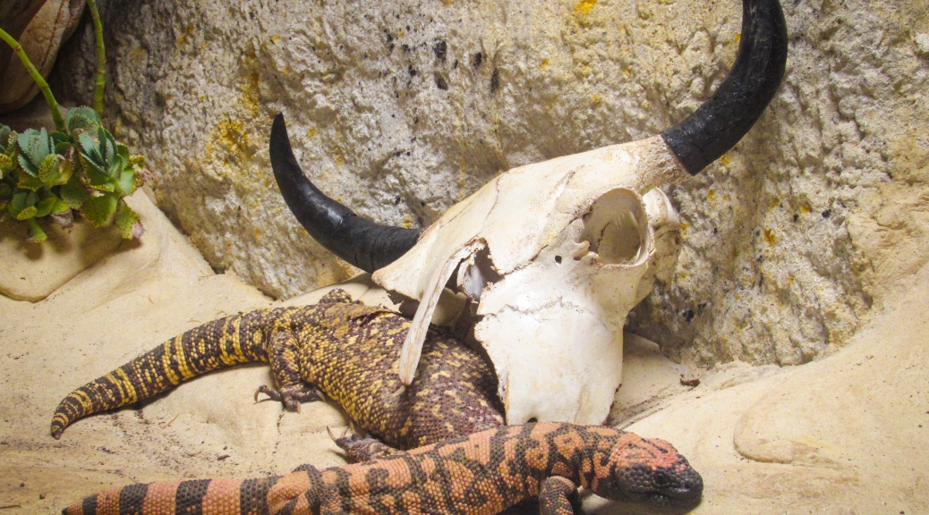
MONSTERS OF THE NEW WORLD
The most famous venomous lizards are the anguimorphs of the Helodermatidae family. From their discovery it was known that these lizards where venomous, as they present a pair of venomous glands in their lower jaws and various pairs of grooved teeth similar to those of venomous snakes with which they inject venom.
Helodermatid skull, in which we can see the sharp teeth with which they inject their venom. Image from Heloderma.net.
The helodermatis are carnivorous animals which feed on small mammals, birds, wall lizards, amphibians, invertebrates, eggs and carrion. Considering its generalist diet and that their prey are pretty defenceless, it is thought that venom evolved in these reptiles as a predator deterrent method, not as a hunting strategy.
Photo by Walknboston of a Gila monster (Heloderma suspectum), in which we can see its black and yellow coloration, with which it warns its predators about its toxicity (aposematic coloration).
The Gila monster and the beaded lizard (Heloderma horridum) are slow animals which aren’t really dangerous to human beings. Yet their raising popularity as exotic pets has ended with some bite cases. The bite of a Gila monster causes some serious and burning pain, local edema, weakness, dizziness and nausea. Even if heavy bleeding is usually associated with bites, this isn’t due to some sort of anticoagulant substance but to the helodermatid’s sharp teeth and to the fact that to inject the venom they must chew their aggressor strongly , causing deep lacerations.
THE BEARDED DRAGON
The saurians of the genus Pogona are iguanians of the Agamidae family. These Australian reptiles are known as bearded dragons for the spines that they present on their throats. Even though they are adapted to live in arid places, the environmental temperature can affect the sex of their offspring.
Photo of an eastern bearded dragon in which we can see its yellow coloured mouth. Could it be that this coloration is indicating anything? Photo by Matt.
Bearded dragons are inoffensive animals, but there’s one species with a secret weapon. The eastern bearded dragon (Pogona barbata) is a venomous lizard but, while the rest of venomous reptiles only have one pair of venomous glands, the eastern bearded dragon has two pairs: two in its upper jaw and two in its lower jaw.
Transversal section of the mouth of an eastern bearded dragon, in which we can see the incipient venomous glands both in its upper jaw (mxivg) and its lower jaw (mnivg). Image extracted from Fry, Vidal et al.
The venom they produce isn’t really strong (in human beings it only causes a minor swelling) and the glands are considered vestigial. Yet, the Toxicofera theory argues that the glands of the bearded dragon show us the primitive form which the first toxicoferan reptile would have presented, with two pairs of venom glands instead of a single pair like most current venomous reptiles.
THE BIG MONITORS
Everyone has heard about monitor lizards (anguimorphs of the Varanidae family). There are hundreds of documentaries about the Komodo dragon in which we are told that these animals have so many bacteria in their mouths that their bites inflict an infection, deadly enough to kill an adult bull. Yet recent studies have shown that the monitor’s poor buccal hygiene is not what causes the death of their victims.
Perente or perentie (Varanus giganteus) a typical varanid, with long neck, strong legs, active metabolism and developed senses. Photo by Bernard Dupont.
Even if there are three frugivorous species, the rest are obligate carnivores. It has always been said that the mouth’s bacteria of the monitors is what causes the death of their prey, even if there isn’t any studies which prove it. In fact, in many studies it has been seen that the monitor’s saliva isn’t very different from that of other herbivorous reptiles.
Photo in which we see the feared monitor’s saliva, specifically from an Asian water monitor (Varanus salvator). Image by Lip Kee.
In a study, it was demonstrated that various species of monitor lizards present venom glands in their lower jaws. These glands are among the most complex venomous glands known of all reptiles. In the case of the Komodo dragon, these are compound glands with a larger posterior compartment and five smaller anterior compartments. These compartments have ducts that carry the venom between the teeth.
Even if varanids are closely related to snakes (they share, for example, a bifid tongue), these don’t present the snakes’ characteristic grooves in their teeth. This is due to the fact that instead of injecting the venom directly, monitor lizards use their serrated teeth to open a deep wound in their prey, through which the venom will enter the organism.
Skull of megalania (Varanus priscus) in which we can see the teeth without gooves. This extinct monitor with more than 5 metres long, was the largest venomous animal known. Photo by Steven G. Johnson.
The utility of the venom for the predatory monitors is also supported by the large quantities of venom that they produce. In constrictor snakes that don’t utilise venom, the genes which codify the synthesis of venom are atrophied because of the great amount of energy required to produce it. Monitors, instead, secrete lots of venom with the slightest stimulation of their glands. This venom contains anticoagulant compounds which prevent the wound to close and also produces a cardiovascular shock in the animal by lowering the blood pressure.
A group of Komodo dragons (Varanus komodoensis) feeding on a recently killed pig. Image extracted from Bull, Jessop et al.
Even if we still don’t know for sure if the common ancestor of all these animals was venomous, nor if venom appeared independently in the different families, the relationship between the different members of the clade Toxicofera has been supported by posterior phylogenetic analyses. What we know is that venom is an extremely powerful weapon in the struggle for survival and that, even if snakes are the most numerous venomous reptiles, many other squamate species have been benefiting from the use of toxins, both for self-defence and to subjugate their prey.
REFERENCES
The following sources have been used during the elaboration of this entry:
- Halliday & Adler (2007). La gran enciclopedia de los Anfibios y Reptiles. Editorial Libsa.
- http://www.nature.com/nature/journal/v439/n7076/full/nature04328.html
- http://www.sciencedirect.com/science/article/pii/S0041010114003353
- http://www.leidenuniv.nl/en/researcharchive/index.php3-c=134.htm
- http://www.heloderma.net/en/heloderma.html
- http://journals.plos.org/plosone/article?id=10.1371/journal.pone.0011097
- https://www.ncbi.nlm.nih.gov/pmc/articles/PMC2690028/
- Cover image by Rocío Corzo.


 Diagram of a crocodile egg: 1. eggshell 2. yolk sac 3. yolk (nutrients) 4. vessels 5. amnion 6. chorion 7. air 8. alantois 9. albumin (white of the egg) 10. amniotic sac 11. embryo 12. amniotic fluid. Image by
  Diagram of an amphibian egg: 1. jelly capsule 2. vitelline membrane 3. perivitelline fluid 4. yolk 5. embryo. Image by
  Reconstruction of Solenodonsaurus janenschi, one of the candidates in being the first amniote, which lived around 320-305 million years ago in what is now the Czech Republic. Reconstruction by
  Diagram of an anapsid skull, by
  Diagram of a synapsid skull, by
  Diagram of a diapsid skull, by
  Drawing of the skull of Archaeothyris, which is thougth to be one of the first synapsids that lived around 306 million years ago in Nova Scotia. Drawing by
  Reconstruction of Dimetrodon grandis, one of the better known synapsids, from about 280 million years ago. Reconstruction by
  Evolutionary tree of current vertebrates, in which green color marks the groups previously included inside reptiles. As you can see, the traditional conception of "reptile" includes the ancestors of mammals and excludes birds. Image by
  Shed skin of a rat snake. Photo by
  Photo of a tuatara (Sphenodon punctatus), by
  Photos of some squamates, from left to right and from top to bottom: Green iguana (Iguana iguana, by
  Drawing of the skull of the dinosaur Massospondylus in which we can see the different characteristic openings of diapsid archosaurs. Image by
  Photo of two species of modern arcosaurs: a Nile crocodile (Crocodylus niloticus) and a yellow-billed stork (Mycteria ibis). Photo by
  Skeleton of the extinct tortoise Meiolania platyceps which lived in New Caledonia until 3000 years ago. In this photo it can be seen the compact cranium without openings. Photo by
  Individual leopard tortoise (Stigmochelys pardalis) from Tanzania. Photo by

