Arxiu d'etiquetes: third eyelid

Check the evolution in your own body

42% of the US population and 11.5% of the Spanish people do not believe in evolution. However, there are different evidence that Darwin was right, some of them in your own body. Have you had your appendix or wisdom teeth removed? Find out in this post which vestigial organs you have inherited from your ancestors.

WHAT ARE VESTIGIAL STRUCTURES?

Vestigial structures (often called organs althouth they are not organs properly) are body parts that have been reduced or have lost its original function during the evolution of a species. They can be found in many animals, including humans.

Esqueleto de orca en el que se observan vestigios de las extremidades traseras. Foto: Patrick Gries
Orca skeleton in which vestiges of the hind limbs can be seen. This is a proof of its terrestrial origins. Photo: Patrick Gries

Vestigial structures were fully functional in the ancestors of these species (and in the homologous structures of other existing species), but currently its function is practically useless or it has changed. For example, the second pair of flying wings in some insects such as flies have lost their function and they have been reduced to balance organs (halteres). If you want to know more about the evolution of flight in insects click here.

Besides physical structures, vestigial features can also manifest itself in behavior or biochemistry processes.

WHY ARE THEY  EVIDENCE OF EVOLUTION?

Natural selection acts on species favoring features that increase their survival and eliminating the ones with no benefits, for example when changes appear in the habitat. Individuals with unfavorable characteristics will die or will breed less and that feature will be removed after some generations, while favorable traits will remain as their carriers can pass them to the next generation.

Sometimes there are features that are neither favorable nor unfavorable, so they continue appearing in the next generations. But all has a cost structure (energy, risk to become infected, develop tumors…), so selective pressure continues acting to eliminate something that is not conducive to the success of the species. This is the case of vestigial structures, which “take longer” disappear throughout evolution. Their existence reveal that in the past these structures had an important role in our ancestors.

FIND YOUR VESTIGIAL TRAITS

THE NICTITATING MEMBRANE

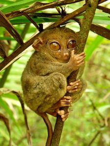
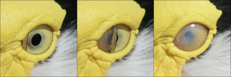
We talked about it in How animals see the world. The third eyelid is a transparent or translucent membrane that protects and moisten the eye without losing visibility. It is common in amphibians, reptiles and birds. Among primates, it is only functional in lemurs and lorises.

membrana nictitante, nictitating membrane
Nictitating membrane or third eyelid of a masked lapwing (Vanellus miles). Photo: Toby Hudson

In humans the plica semilunaris is a remnant of the nictitating membrane. Obviously we can not move it but still has some lacrimal drainage function and helps on the eye movement (Dartt, 2006).

Plica semilunaris (pliegue semilunar). Foto: desconocido
Plica semilunaris. Photo: unknown

DARWIN’S TUBERCLE AND EAR MUSCLES

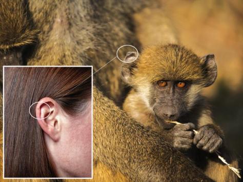
10% of the population has a thickening in the ear, a vestige of the common pointy ear in primates. This structure is called Darwin’s tubercle and has no function.

Variabilidad del Tubérculo de Darwin en la punta de la oreja (0= ausente). Puede presentarse en otras zonas del pabellón auditivo: ver publicación.
Variability of Darwin’s tubercle at the top of the ear (0 = absent).  Credit.
Comparación entre la oreja de un macaco y la nuestra. Fuente
Comparison between the ear of a yellow baboon (Papio cynocephalus) and ours. Credit

Also, primates (and other mammals) have mobile ears to lead the pinna toward the sound source: surely you have noticed it in your house dog or house cat. Humans (and chimps) no longer have that great mobility, although some people may move slightly pinna. It has been proven with electrodes these muscles are excited when we perceive a sound that comes from a particular direction (2002).

Auricular muscles responsible of movement of the pinna. Credit

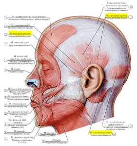
The occipitofrontalis muscle has lost its function to prevent the head from falling, but participates in facial expression.

PALMARIS LONGUS MUSCLE

16% of Caucasians do not have this muscle on the wrist, neither 31% of nigerian people neither 4,6% of chinese people. It can even appear in one arm and not in the other or be double.

It is believed that this muscle actively participated in the arboreal locomotion of our ancestors, but currently has no function, because it does not provide more grip strength. This muscle is longer in completely arboreal primates (like lemurs) and shorter in land primates, like gorillas (reference).

And do you have it or not? Try it: join your thumb and pinky and raise your hand slightly.

mireia querol, mireia querol rovira, palmaris longus, musculo palmar largo, tendon
I have two palmaris longus in the left arm and one on the right. Photo: Mireia Querol

WISDOM TEETH

35% of people do not have wisdom teeth or third molar. In the rest, its appearance is usually painful and removal is necessary.

Yo no tengo el tercer molar. Foto: Mireia Querol Rovira
I don’t have the third molar. Photo: Mireia Querol Rovira

Our hominin ancestors had them, much bigger than ours. A recent research explains that when a tooth develops, emits signals that determine the size of the neighboring teeth. Reducing the mandible dentition and the other along evolution has resulted in reduced molars (and eventually the disappearance of the third).

Comparativa entre la dentición de un chimpancé, Australopithecus afarensis y Homo sapiens. Fuente
Comparison between the dentition of a chimpanzee, Australopithecus afarensis and Homo sapiens. Look at the reduction of the last three molars between afarensis and sapiens, Credit

THE TAILBONE

If you touch your spine till the end, you will reach the coccyx or tailbone. It is three to five fused vertebrae, vestige of the tail of our primate ancestors. In fact, when we were in the womb, in the early stages of embryo development a 10-12 tail vertebrae formation is observed.

Distintos estados en el desarrollo embrionmario humano y comparación con otras especies. Créditos en la imagen
Different stages in human embryonic development (1 to 8) and comparison with other species. Credits in the image.

Subsequently it is reabsorbed, but not in all cases: it has been reported 40 newborns with a tail.

Neonato nacido con cola. Una mutación ha evitado la inhibición del crecimiento de la cola durante la gestación. Fuente
Infant born with tail. A mutation has prevented the growth inhibition of the tail during pregnancy. Credit

Although we have no tail, currently these bones serve as anchors of some pelvic muscles.

mireia querol, mireia querol rovira, coxis, sacro, sacrum, tailbone, rabadilla
Tailbone position. Photo: Mireia Querol Rovira

SUPERNUMERARY NIPPLES (POLYTHELIA)

It is estimated that up to 5% of the world population has more than two nipples. These “extra” nipples can be presented in different ways so sometimes are confused with freckles or moles. They are located in the mammillary line (from the axilla to the groin), exactly in the same position as other mammals with more than two breasts (observe your house dog, for example). Usually the number of breasts corresponds to the average of offspring that has a mammal, so extra nipples would be a vestige from when our ancestors had more offspring per birth. Usual is 3 nipples, but has been documented a case of up to 8 nipples in a person.

Pezón suplementario debajo del principal. Fuente
Additional nipple below the main one. Credit

FIND YOUR VESTIGIAL REFLEXES AND BEHAVIOURS

PALMAR AND FOOT SOLE GRASP REFLEX

Surely you’ve experienced that if you bring anything into the hands of a baby, automatically he grabs it with such a force that would be able to hold his own weight. This reflex disappears at 3-4 months of age and is a remnant of our arboreal past and the way to grab the hair of the mother, as with the other current primates. Watch the next video in 1934 on a study of twins (minute 0:34):

On the feet there is also a reflex of trying to grab something when the foot of a baby is touched. It disappears at 9 months of age.

By the way, have you noticed how easily children climb on any handrails or higher zones in a playground?

GOOSEBUMPS

Cold, stress or intense emotion (eg, listening to some music) causes the piloerector muscle to raise the hair giving the skin the appearance of a plucked chicken. It is an involuntary reflex in which some hormones, like adrenaline (which is released in the mentioned situations) are involved. What utility had this to our ancestors and has in modern mammals?

  • Increasing the space between the skin and the external surface, so that hot air trapped between hair helps on maintaining temperature.
  • Looking bigger to scare off potential predators or competitors.
Chimpancé con el pelo erizado durante un display antes de un conflicto. Foto: Chimpanzee Sanctuary Northwest
Chimpanzee with hair bristling in a display before a conflict. Photo: Chimpanzee Sanctuary Northwest

Obviously we have lost hair in most parts of the body, so although we retain the reflex, it has no use to us or to keep warm or to ward off predators. The hair has been preserved abundantly in areas where protection is necessary or due to sexual selection (head, eyebrows, eyelashes, beard, pubis…), but in general, can also be considered a vestigial structure.

There are more vestigial structures but in this post we have focused on the most observable. In future posts we will discuss other internal structures, like the famous appendix or vomeronasal organ.

REFERENCES

How animals see the world?

Have you ever heard that dogs see in black and white? Or that cats can see in the dark? Why we have our eyes in front of the face? And why goats have an horizontal pupil? This article will answer these and other questions about the eyes and vision, focusing on mammals.

HOW IMAGES ARE FORMED?

The eyes are the receptors responsible for capturing light and sending the signal through the optic nerve to the brain, which make the interpretation. Light is an electromagnetic wave as infrared, ultraviolet, X rays, microwaves, etc. In this post we will refer to visible light, that is, the part of the spectrum that can perceive humans and most mammals.

eye parts
Parts of the eye. Source

Basically, the light passes through the pupil. It can regulate the amount of light thanks to the muscles associated with iris (which gives color to the eye). The lens focuses the objects. The image is projected inverted in the retina, to be sent as an electrical signal to the brain.

WHY DO WE SEE IN COLOR ?

In the retina there are two main types of photoreceptor cells: cons and rods. The main differences are:

RODS
  • More sensitive in a few light conditions
  • No color vision
  • Motion-sensitive
  • Less image detail
CONES
  • Activated under conditions of high light
  • Color vision
  • Contrast-sensitive
  • High image detail

That’s why in low light, vertebrates see in black and white and the image is not clear, since the rods are activated at maximum but the cones are inactive. Some primates have three different kinds of cones (trichromatic vision), which correspond to the red, green and blue colour (RGB). Some primates and other animals have monochromatic vision (they only have one type of cone) or dichromatic (two). Some animals have tetrachromic vision, like birds.

The cones are sensitive to different wavelengths, different colors. Photo taken from Colombian Primatological Association

Generalizing a lot, diurnal vertebrates have more cones than rods and nocturnal ones have more rods than cones, allowing them to see better in the dark. But they can really see in the dark?

SEEING IN THE DARK

In total absence of light it is impossible to see, although some animals can detect other radiation such as infrared (snakes) or ultraviolet (bees). In addition to the relation between rods and cones, other factors that improve vision in low light conditions are:

THE CORNEA

The bigger the eye and the cornea, the better use of light. The mammal with the greatest cornea in relation to the eye is the Philippine tarsier (Carlito syrichta ) a nightlife primate.

Philippines’ tarsier (photo: Yeo Kok Leng)

THE PUPIL

Another way to take advantadge of few light conditions is increasing the size of the pupil. According to the shape of it, the control of incoming light is more precise: it is the case of many cats. Compared with a round pupil, the elongated one opens and closes sideways and according to the position of the eyelid, pupil surface exposed to light can be controlled better.

The felines with vertical pupil can open it horizontally and control better the entry light than with a circular pupil. Image of an unknown author, adapted from Aquàrium-Liège Museum


THE TAPETUM LUCIDUM

Cats, dogs, bats, horses, whales, crocodiles, cattle and some nocturnal primates have in the retina or behind it a bright layer called tapetum lucidum, which increases up to 6 times the light gathering ability compared to humans. As if it were a mirror, the tapetum lucidum reflects the light reaching the eye to return back to the retina and harness light to the maximum.

Reflection of light due to the tapetum lucidum. Image taken from Exclusively cats

The tapetum lucidum is responsible for cat’s eyes appearing to glow in the dark and cat and dog’s pupils shine in blue/green when light falls upon the eye.

Tapetum lucidum shining on a dog. Photo Mireia Querol

WHY SOME ANIMALS HAVE THE EYES IN FRONT OF THE FACE WHILE OTHERS HAVE THEM ON THE SIDES ?

The position of the eye in mammals can be frontal, like a cat, or in the side, like a rabbit. This means distinct advantages:

  • Binocular vision (stereoscopic): allows a good estimation of distance, but the field of view is smaller. A 3D image is generated. It is typical of carnivores that should focus attention to their prey or primates that should calculate the distance between the branches.
  • Side vision (peripheral): allows each eye to send a different signals to the brain, so it is easier to notice their surroundings having a field of view of about 360 degrees. It is typical of herbivores, which must pay attention to the presence of potential predators .

    Visual field of a cat and a horse. The blind area is smaller in hervibores. Source: Sjaastad, Sand and O. Hove K. Photo taken from Eye Opener

WHY GOATS HAVE AN HORIZONTAL PUPIL?

In addition to the position of the eyes, the shape of the pupil is also related if you are a predator or a prey. Goats or horses have horizontal pupils, while cats like the margay have it vertical.

Pupil of a goat (horizontal) and a cat (vertical) Photo: Wikimedia Commons

Banks  says that “to calculate distances predators basis on stereoscopic vision (works better with a small pupil) and sharpness (works best with a larger one). Vertical pupils are small horizontally and large vertically”.

In the case of terrestrial prey attacked by predators, the tendency of the pupil is being horizontally because “can gather more light and and also reduces the sunlight, which could dazzle “. Exceptions such as rabbits or mice with a circular pupil, are because they have to pay attention also to the sky, from where a bird of prey can attack.

WHAT IS THE THIRD EYELID?

Some animals have the nictitating membrane (“third eyelid”), a transparent or translucent membrane that is used to protect and moisten the eye without losing visibility. Camels, seals and polar bears have it complete, whereas in other mammals, such as dogs or humans remains only reduced.

Nictitating membrane in a feline. Photo by Editor B

IS IT TRUE THAT DOGS AND BULLS SEE IN BLACK AND WHITE ?

Actually dogs and cats are able to detect colors, particularly gray, yellow and blue in softer tones. Cats may be able to perceive more colours.

Visible spectrum by a dog and a human. Source

In the case of bulls, it is also spread the myth that rage against the red colour or see in black and white. Actually bulls have dichromatic vision, like most diurnal mammals, since they only have blue and green cones. Therefore, they can’t see red, but it does not mean they see in black and white.

AND OTHER MAMMALS?

Horses see in blue and red tones. Most rodents see in black and white. Most species of the family of goats, sheeps and bulls see from green to violet. In addition, recent studies indicate that many mammals (especially nocturnal ones), contrary to what was believed, also can perceive ultraviolet radiation: rats and mice, reindeer, possibly cats and dogs, cows, pigs, ferrets, okapi…

We finish with a BuzzFeed video with the simulation of vision of some animals. If you have more questions about animal’s vision leave it in the comments!

REFERENCES